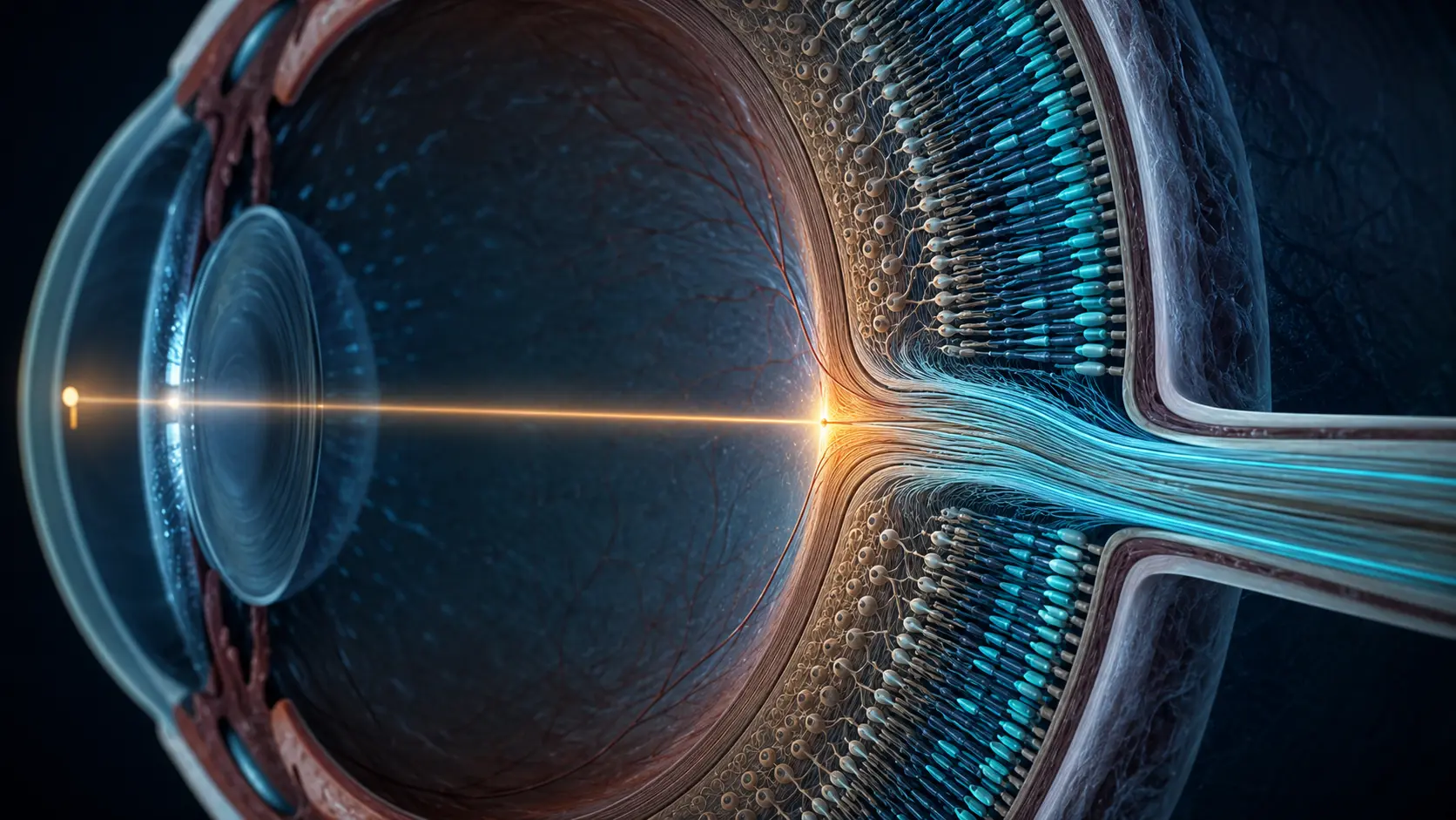
Adaptation changes the balance between color channels.
Cone photoreceptors respond across overlapping wavelength ranges. Downstream neurons compare those signals in opponent channels, including red–green and blue–yellow relationships. Prolonged stimulation shifts the sensitivity of part of that system.
When the strong color is replaced by a neutral field, the adapted channel contributes less than its opponent. The temporary imbalance can be perceived as a complementary afterimage. It fades as sensitivity returns toward baseline and the eyes move across the scene.
The exact hue and strength vary with screen calibration, brightness, fixation, viewing time, surrounding colors and individual sensitivity. Seeing a weak color—or no clear color in one attempt—does not by itself indicate a vision problem. Repeating the test after a brief rest may produce a different result.
Negative afterimages are commonly explained with both receptor adaptation and opponent processing. The familiar red–green and blue–yellow account is useful, but modern color science does not reduce every afterimage to one simple pair of exhausted sensors; retinal and later neural stages contribute together.
Why it matters
Afterimages reveal that color is constructed from comparisons and adaptation, not copied directly from a wavelength onto a mental screen.
The demonstration also separates a common short-lived visual effect from persistent or unexplained visual changes. A normal afterimage fades; ongoing symptoms deserve professional advice.
Because the test depends on a display and subjective report, it cannot measure color deficiency, retinal function or neurological health. Standardized clinical tests use calibrated targets and professional interpretation.
Research behind the test
These references support the mechanism. Your browser result is not a standardized measurement.
01