Quick answer
Your lungs defend against infection through several overlapping layers. Mucus traps many inhaled microbes and particles, while beating cilia move that mucus toward the throat. Airway lining cells produce antimicrobial molecules and detect danger. In the alveoli, resident macrophages engulf material that reaches the gas-exchange surface. If those defenses are exceeded, epithelial and immune cells release signals that recruit neutrophils, monocytes and lymphocytes from the blood. This response can control infection, but too much inflammation can also interfere with breathing.
You inhale thousands of litres of air each day. Along with oxygen come dust, droplets, pollen, microbes and fragments of the environment. Yet the gas-exchange surface at the end of the airways must remain exceptionally thin—only a small barrier can separate air from blood without slowing oxygen transfer.
The lungs solve that conflict with defenses arranged by location. The nose and branching airways trap and transport material upward. The smallest air spaces rely more heavily on resident immune cells and chemical signals. Most encounters are handled quietly; inflammation is a powerful backup, not the default response to every particle.
The airways run a moving barrier
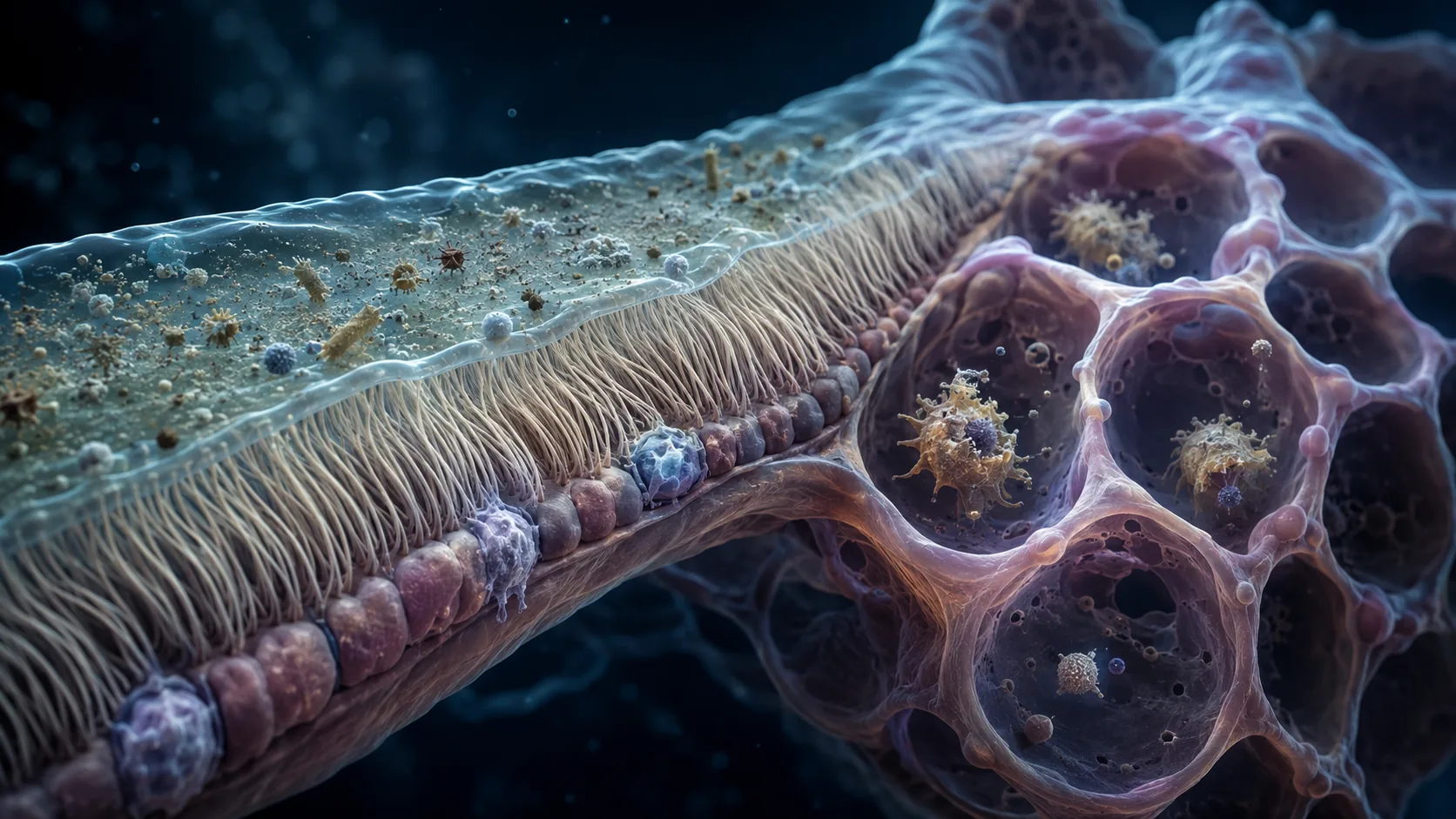
The conducting airways are lined by an epithelium containing mucus-producing cells and ciliated cells. Mucins give mucus its gel-like properties, allowing it to catch particles that settle on the airway surface. Beneath that layer sits a thinner fluid zone in which microscopic cilia can beat.
Coordinated ciliary motion pushes the mucus layer upward toward the throat. The material can then be swallowed or coughed out. This mucociliary escalator removes a potential threat physically, often before immune cells need to launch a damaging inflammatory response.
The system depends on the right hydration, mucus consistency and ciliary movement. Smoke can injure cilia, while diseases that produce abnormally dehydrated mucus or defective ciliary motion impair clearance. A cough adds a faster mechanical route when mucus or irritation activates airway sensors.
Airway lining cells are active sentinels
The epithelium is not inert packaging. Airway cells express pattern-recognition receptors that respond to molecular features associated with microbes or tissue damage. They also produce antimicrobial proteins, mucus components and signaling molecules that shape the local response.
This surface has to discriminate. The respiratory tract meets harmless material continuously, so an aggressive reaction to every exposure would injure the very tissue needed for gas exchange. Barrier integrity, controlled signaling and clearance work together to maintain tolerance while preserving the ability to escalate.
When epithelial cells detect a meaningful threat, they can release cytokines and chemokines. These chemical gradients help activate nearby cells and guide additional immune cells toward the affected tissue. The response is therefore organized locally before it becomes a whole-body event such as fever.
Alveolar macrophages patrol the deepest air spaces
Mucus would be a poor coating for the alveoli because it would obstruct the thin, moist surface required for oxygen and carbon dioxide exchange. Instead, alveolar macrophages sit within the air spaces and sample particles that escape the upper defenses. They can engulf microbes, dust and dying cells through phagocytosis.
In a healthy lung these macrophages also restrain unnecessary inflammation. That balancing role is essential: a large influx of fluid and immune cells may help fight infection, but it can thicken the gas-exchange barrier. Protection requires enough force to control the threat without turning the alveolus into a battlefield that cannot exchange gases efficiently.
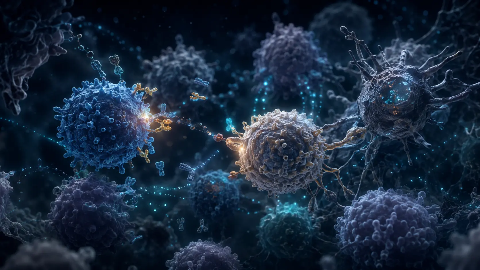
If resident defenses are not enough, macrophages and epithelial cells recruit neutrophils and monocytes from the bloodstream. Dendritic cells can carry antigen information toward draining lymph nodes, where T and B lymphocytes may be activated. Innate defense buys time while a more specific adaptive response develops.
What happens when a pathogen gets past the barriers
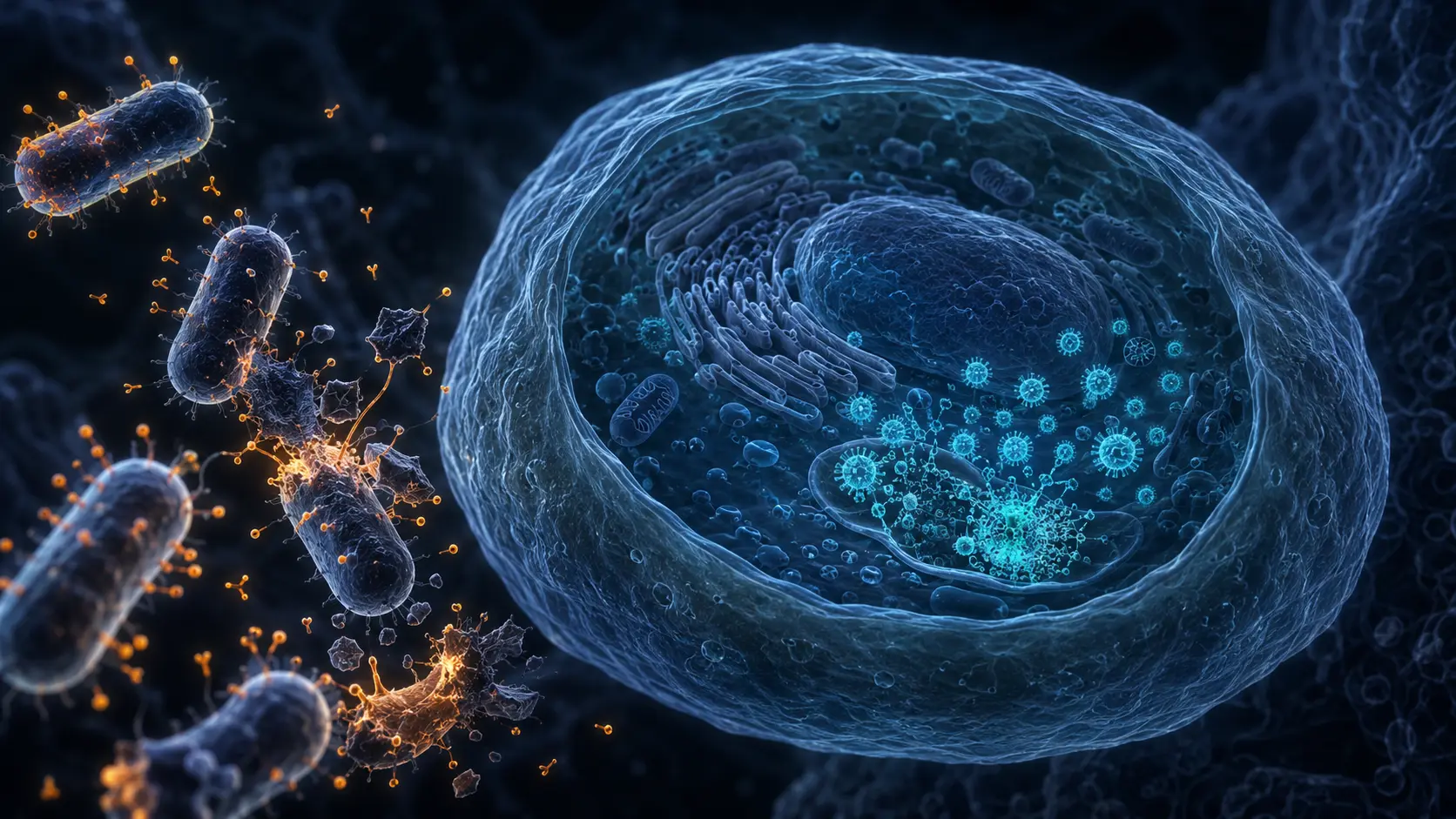
Pathogens succeed in different ways. Some attach to airway cells despite mucus, some damage cilia, and some replicate inside epithelial cells. A high dose or an impaired defense system can also shift the balance. The resulting inflammation produces symptoms such as cough and increased mucus, but symptoms alone cannot identify whether the cause is viral, bacterial or something noninfectious.
Vaccination can prepare adaptive immune memory so specific B and T cells respond more quickly to particular pathogens. It does not replace mucus, cilia or macrophages; it adds a faster targeted layer to the same defense network. Likewise, antibiotics only treat susceptible bacteria and do not strengthen the lung's physical barriers or treat viruses.
Breathlessness, chest pain, confusion, blue or gray lips, or rapidly worsening symptoms require medical assessment. The mechanism explains normal defenses; it cannot diagnose an infection or measure its severity at home.
From inhaled particle to immune response
Location determines the first responder. In larger airways, mucus catches material and cilia move it upward. In the alveoli, resident macrophages engulf and assess what arrives. Barrier cells and immune cells exchange signals continuously.
When local control fails, chemokines recruit circulating leukocytes and antigen reaches lymph nodes. A specific adaptive response can then expand, while regulatory pathways work to limit collateral damage and restore the thin gas-exchange surface.
A respiratory review describes the partnership between mucins, mucociliary clearance and macrophages.
A current barrier review maps the physiological and immunological defenses of the lung.
Try it yourself
Notice clearance without trying to provoke it.
- Notice that a mild cough can move material upward rather than deeper into the chest.
- Compare nasal breathing with mouth breathing during normal rest; the nose adds filtering, warming and humidification.
- Avoid smoke and deliberate exposure to dust, aerosols or extreme cold as an experiment.
This is not a lung-function or infection test. Persistent cough, breathing difficulty or concerning symptoms need qualified medical advice.
Why it matters
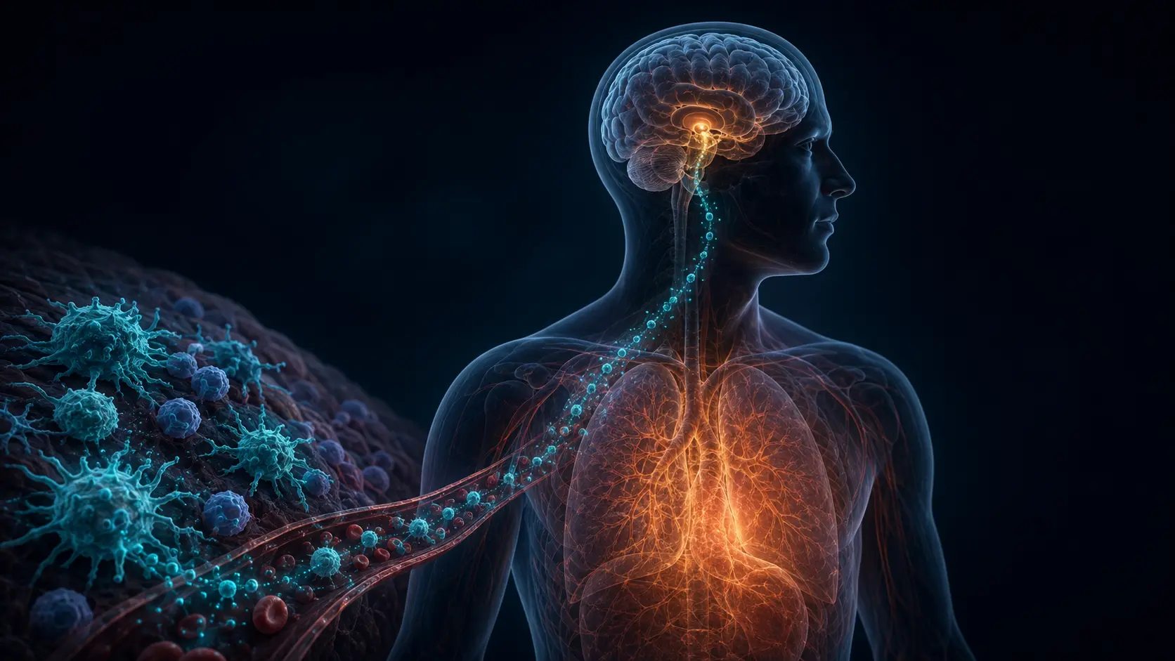
The respiratory and immune systems are not separate departments. Physical transport, epithelial sensing, resident immune cells, blood recruitment and lymph-node activation form one connected defense route.
The mechanism also explains why more inflammation is not always better. The lungs must destroy threats while preserving an extremely delicate surface for gas exchange.
Your lungs defend themselves in layers.
Mucus and cilia remove many threats mechanically, macrophages patrol the alveoli, and recruited immune cells provide backup when local control is not enough.
Research behind this story
We link to the primary study or an authoritative indexed review wherever possible. Caveats in the text reflect the limits of that evidence.
01