Quick answer
Eye floaters appear when tiny clumps, fibres or cells within the vitreous gel scatter or block light and cast moving shadows on the retina. They are easiest to notice against a bright, uniform background and seem to drift because the vitreous continues moving after the eye turns. Age-related vitreous changes are common, and many longstanding floaters are harmless. A sudden shower of new floaters, flashes of light, a curtain-like shadow or missing vision can signal a retinal tear or detachment and needs urgent dilated eye examination.
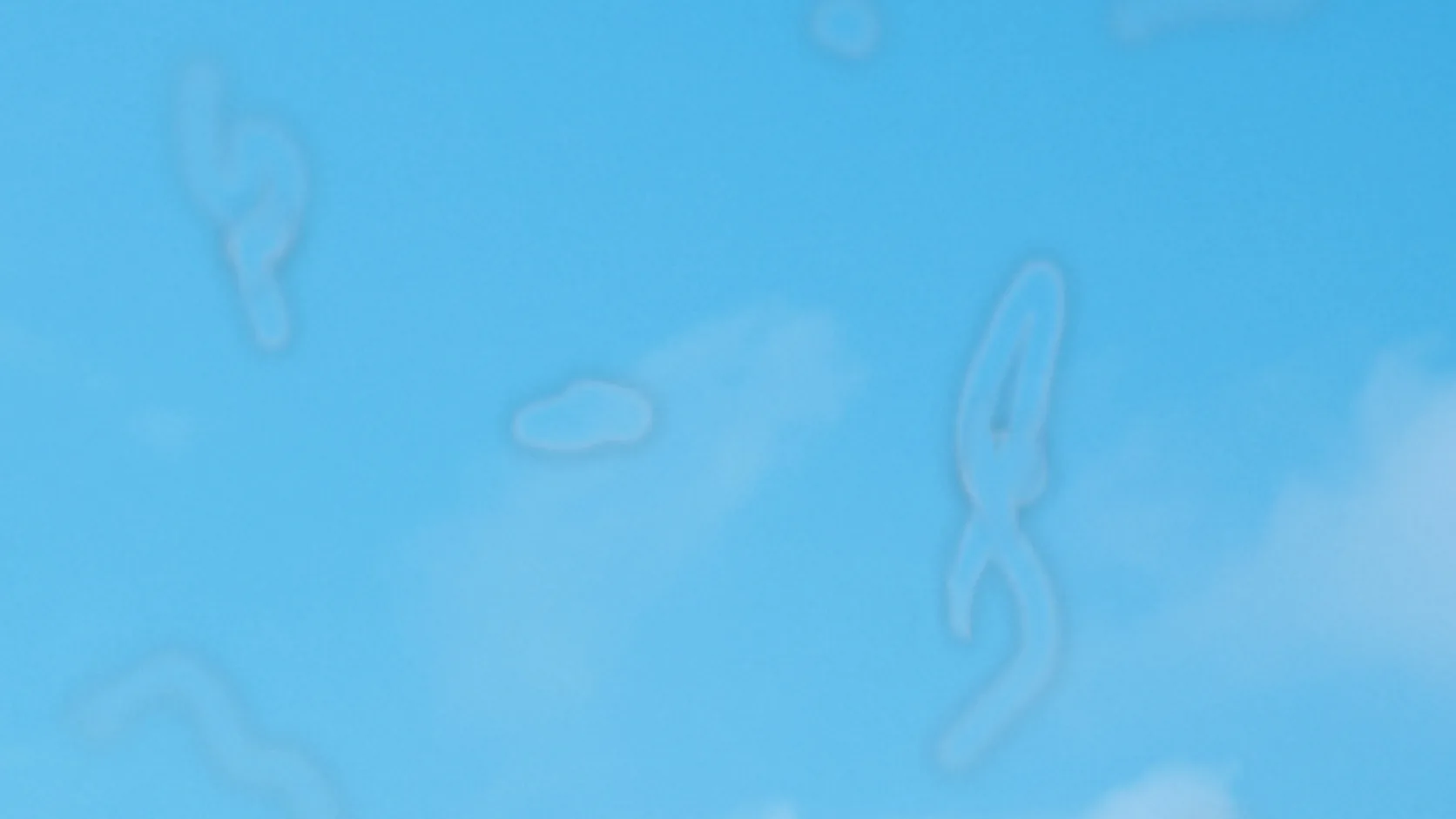
Look at a clear sky or white wall and a transparent thread may drift into view. Try to stare directly at it and it slips away, then glides back after the eye stops moving.
The object is not on the surface of the eye and it is not floating in the outside world. Most floaters are small structures within the vitreous—the clear gel filling the space between lens and retina. You see their shadows, not the structures themselves.
The shadow begins inside the vitreous
The vitreous body is mostly water held in a transparent network of collagen and hyaluronan. In youth that structure is relatively uniform. With age, parts of the gel liquefy and collagen fibres can aggregate, creating small regions that interact with incoming light.
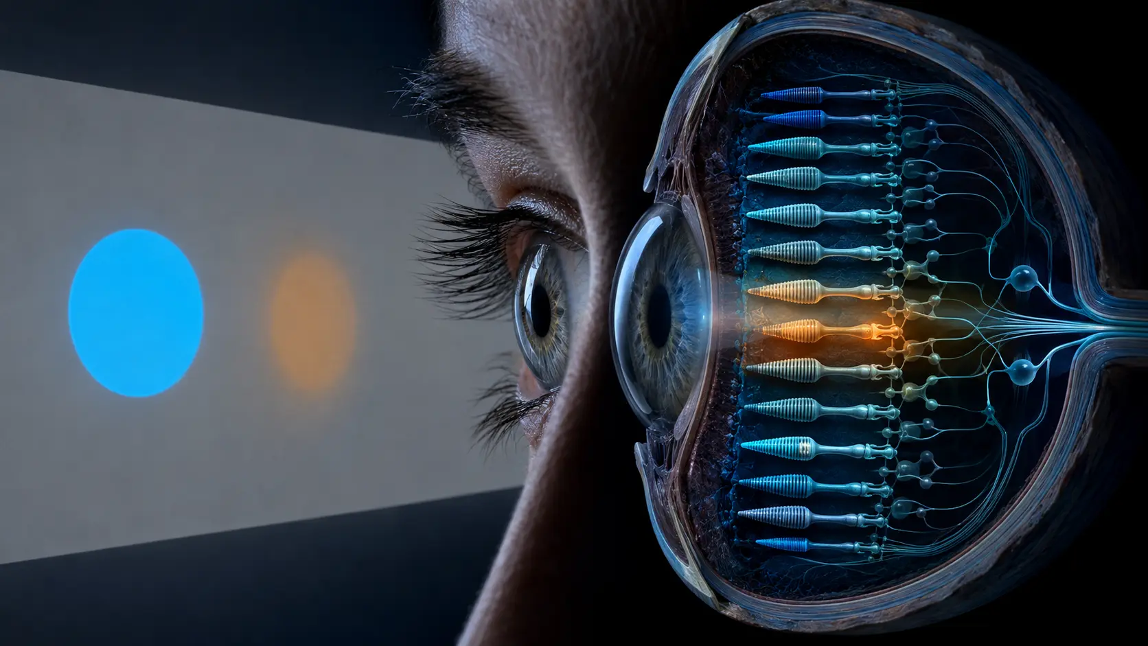
When an opacity lies in the light path, it casts a defocused shadow onto the retina. The retina converts that changing light pattern into neural signals, and the visual system interprets the shadow as a spot, strand, cobweb or translucent shape suspended in the visual field.
Floaters are not sharply focused because they sit in front of the retina rather than on it. Their visibility depends on size, density and distance from the retinal surface. Opacities closer to the retina can produce more defined shadows.
Why floaters appear against the sky or a white screen
A bright uniform field gives the retina an even background, so a small shadow creates strong local contrast. In a detailed or dim scene, edges and texture compete with the floater and make it easier for the visual system to ignore.
Turning the eye sets the vitreous in motion. Because the gel has inertia, internal strands lag behind and continue drifting after the eyeball stops. That is why a floater seems to flee when you try to look directly at it and then settles back into view.
Over time, some opacities shift away from the most sensitive visual axis and the brain may reduce attention to a familiar pattern. The floater may still be physically present even when it becomes much less noticeable.
How age changes the vitreous-retina connection
The vitreous is attached to the retina at several points. As it liquefies and contracts with age, it can separate from the back of the eye in a posterior vitreous detachment. This is common later in life and may create a new ring, strands or a sudden increase in floaters.
Most posterior vitreous detachments do not cause lasting harm, but traction can sometimes tear the retina. Fluid can then pass through the tear and separate the retina from the tissue beneath it. Because retinal detachment can threaten vision without causing pain, symptom timing matters.
Myopia, prior cataract surgery, eye trauma and some inflammatory or retinal conditions can alter risk. Only a dilated examination can determine whether sudden symptoms come from uncomplicated vitreous change or a retinal problem.
When new floaters need urgent attention
A few stable floaters that have been present for a long time are common. A sudden shower of new spots, repeated flashes at the side of vision, a dark curtain or shadow, or new missing or blurred vision is different. These can be warning signs of a retinal tear, bleeding or detachment.
An urgent dilated eye examination allows an eye-care professional to inspect the peripheral retina. Waiting to see whether a curtain-like shadow clears can risk permanent vision loss if the retina is detaching.
Rubbing the eye, staring at bright light or trying to chase the floater cannot identify its cause. Online images can explain the optics but cannot replace examination when the pattern changes suddenly.
From vitreous strand to drifting shape
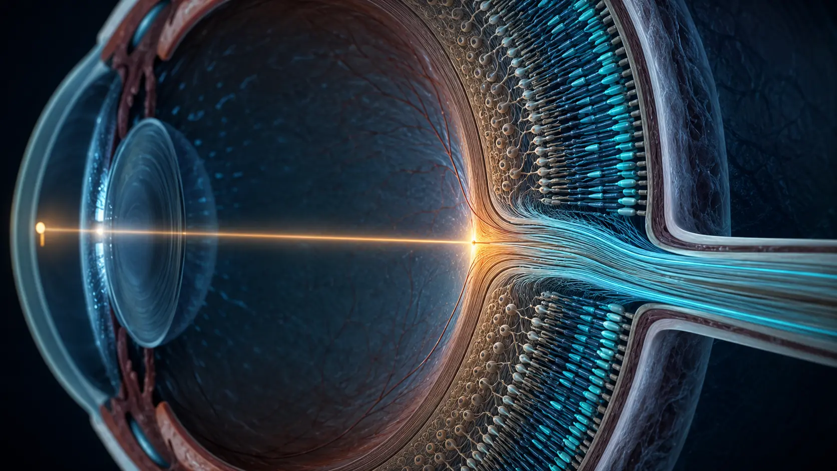
Light enters through the cornea and lens, then passes through the vitreous. A clump or fibre alters part of that beam and projects a soft shadow onto retinal photoreceptors.
Eye movement shifts the gel and the opacity continues to drift. The moving retinal shadow becomes the spot or cobweb you perceive against the external scene.
The National Eye Institute explains how vitreous strands cast new shadows during detachment.
A clinical review details the relationship between posterior vitreous detachment, floaters and retinal tears.
Try it yourself
Use a safe, evenly lit surface.
- Look at a plain light wall or overcast sky—not the sun or another intense light source.
- Move your eyes gently left and right, then stop and notice whether a shape continues drifting.
- Stop the observation and seek care if floaters are suddenly new or accompanied by flashes, a curtain or lost vision.
Never look directly at the sun to see floaters. Sudden warning symptoms require urgent professional assessment.
Why it matters
Floaters reveal the optical path inside your own eye: an internal structure becomes visible only because it changes the light reaching the retina.
The same mechanism also carries a safety lesson. Common longstanding shadows and sudden retinal warning signs can feel similar, so a rapid change should not be dismissed as normal aging.
A floater is an internal opacity seen as a retinal shadow.
Vitreous strands drift with eye movement and cast shadows on the retina; sudden new floaters with flashes or lost vision need urgent examination.
Research behind this story
We link to the primary study or an authoritative indexed review wherever possible. Caveats in the text reflect the limits of that evidence.
01