Quick answer
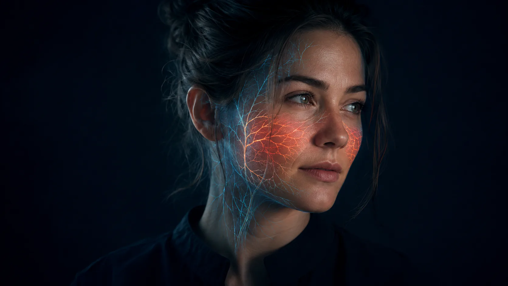
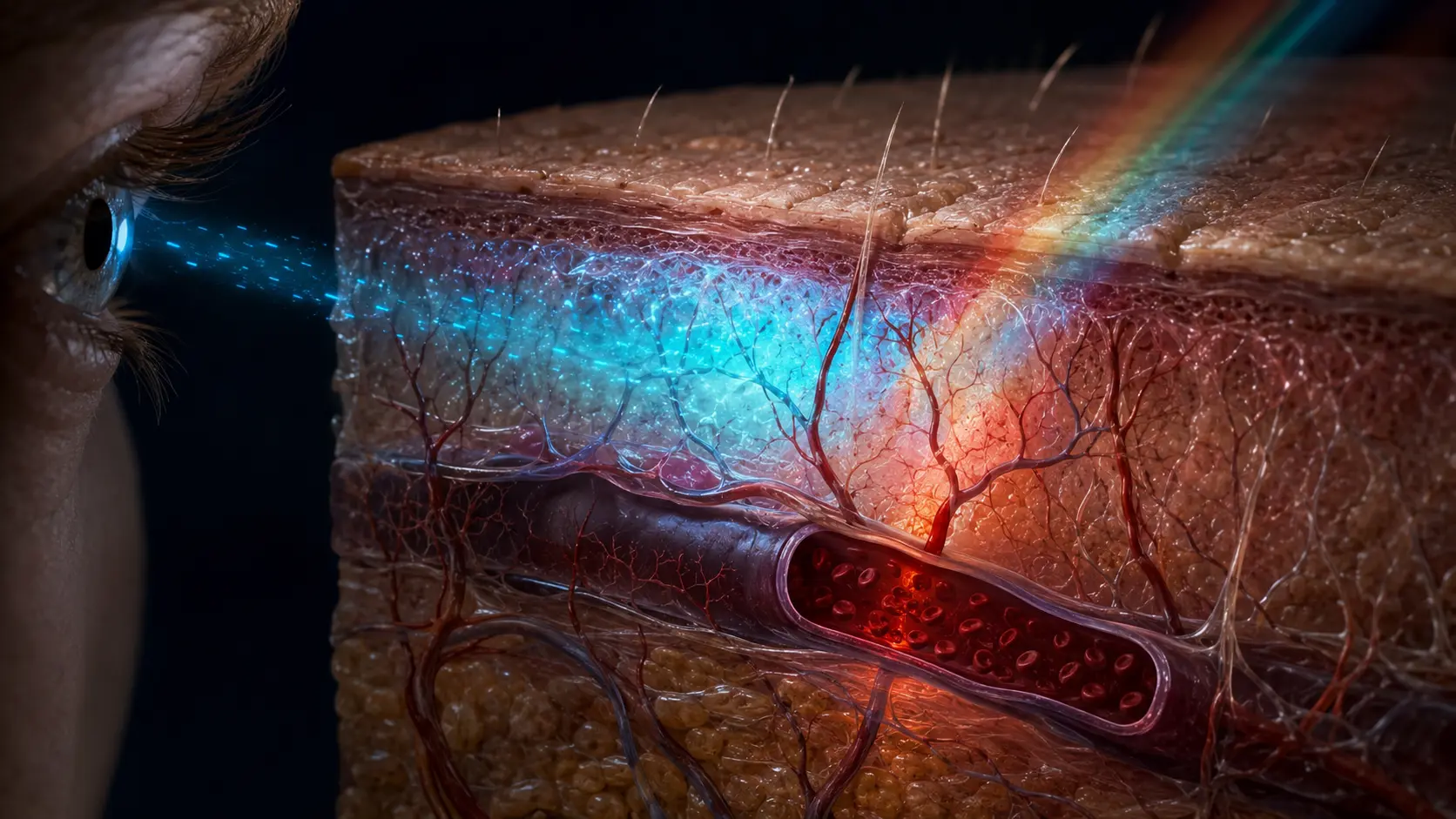
Veins look blue because light must travel through skin and tissue before and after interacting with the vessel. Red wavelengths penetrate more deeply and are absorbed differently by blood and surrounding tissue, while shorter wavelengths are scattered and returned differently to the eye. The combined optical contrast can make a superficial vein appear blue or green even though the venous blood inside it is dark red.
Look at the inside of your wrist and several vessels may appear blue, teal or green. The simple explanation often offered in school is that veins carry blue blood until oxygen reaches it. That explanation is wrong.
Human blood is always some shade of red. Oxygen-rich blood is brighter because of the way oxygen changes hemoglobin; oxygen-poor venous blood is darker. A vein only looks blue when you view it through layers of living tissue and reflected light.
Color is the light that makes it back
White light contains many visible wavelengths. When it reaches skin, some is reflected at the surface, some is absorbed by pigments such as melanin and hemoglobin, and some scatters through cells, collagen and other microscopic structures. The pattern that returns to the eye determines perceived color.
Blood absorbs light in wavelength-dependent ways. Oxygenated and deoxygenated hemoglobin have different spectra, which is why medical instruments can estimate oxygen saturation with carefully chosen red and infrared light. Neither spectrum turns blood blue; a vial of venous blood still appears deep red.
A vessel changes the light returning from a small patch of skin compared with the tissue beside it. Depth, diameter, surrounding fat, skin pigmentation and illumination all influence that contrast. The brain interprets the relative color, so the same vein can look more blue, green or nearly invisible under different conditions.
Why superficial veins are easiest to see
Many arteries run deeper and have thicker muscular walls, while some veins travel close to the skin. A shallow vessel can alter reflected light strongly enough to be visible. As depth increases, repeated scattering softens the contrast and the outline disappears.
Skin color changes the balance because melanin absorbs broadly across visible wavelengths. That can reduce vessel contrast without changing the blood itself. Temperature and pressure also change vein size: warmth can dilate superficial vessels, while raising an arm or pressing the skin can reduce how prominent they look.
Medical diagrams often use red for arteries and blue for veins as a map of flow direction and oxygenation. The convention is useful, but it is symbolic. Pulmonary arteries carry oxygen-poor dark red blood, and pulmonary veins carry oxygen-rich bright red blood—the names describe direction relative to the heart, not color.
Why your veins can look blue, green or more visible
There is no single vein color that should appear on every person. Melanin, the thickness and composition of the tissue above the vessel, vessel depth and the color of the illumination all change the spectrum that reaches the eye. A green cast can emerge from the same optical comparison that produces blue; it does not mean the blood has changed color.
Veins may stand out more after warmth or exercise because superficial vessels widen as the body redistributes heat. They can also look more prominent where skin is thin or subcutaneous fat is limited. Raising a limb changes venous filling, and warm indoor light can shift the apparent hue compared with daylight.
Visible blue veins are usually normal anatomy, not evidence that the blood lacks oxygen. A pulse oximeter estimates arterial oxygen saturation using red and infrared light; looking at vein color cannot do that job. Sudden swelling, pain, redness, warmth, a hard cord or a major one-sided change is a different question and deserves medical assessment rather than an optical explanation.
Blue-vein questions, answered directly
Why do my veins look blue if blood is red? Because the surrounding skin and tissue alter returning light, and the visual system judges the vessel relative to nearby skin. Why are veins not bright red? Venous blood is darker red than arterial blood, and the vessel is being viewed through tissue rather than directly.
Are only veins able to look blue? No. Models show that sufficiently deep arteries can also acquire a bluish appearance, although differences in depth, wall structure and hemoglobin absorption often make superficial veins easier to see. The familiar color code in diagrams is therefore a teaching convention, not a literal photograph of circulation.
How dark red blood becomes a blue-looking line
Light enters the skin, where pigments and tissue remove or redirect different wavelengths. The vessel absorbs part of what reaches it. Light that returns through the same tissue is spectrally altered, and the visual system compares that patch with nearby skin.
The result is a context-dependent blue or green appearance. Cut the optical path—by seeing blood directly rather than through skin—and the familiar red returns.
Biomedical optics reviews explain how hemoglobin absorption and tissue scattering shape vessel appearance.
Controlled vessel phantoms confirm that depth and wavelength change the visible color.
Optical modelling shows that the blue impression reflects a larger relative loss of red light over the vessel.
Try it yourself
Change lighting, not circulation.
- View the same wrist under daylight and a warm indoor light.
- Compare a shallow vein with one that fades deeper into the arm.
- Notice the color relative to surrounding skin rather than naming it in isolation.
Do not tie off, strike or repeatedly press a vein. Changes in skin or vessel appearance that concern you should be assessed medically.
Why it matters
The question corrects a durable misconception about circulation: deoxygenated does not mean oxygen-free, and it never makes human blood blue.
It also reveals how much of seeing is comparison. The eye receives wavelengths, but the brain builds color from illumination, surrounding tissue and neighboring surfaces.
The vein is dark red; the view through skin looks blue.
Wavelength-dependent absorption, scattering and visual contrast transform the returning light. The apparent blue is produced by the path, not the blood.
Research behind this story
We link to the primary study or an authoritative indexed review wherever possible. Caveats in the text reflect the limits of that evidence.
01