Quick answer
A fetus gets oxygen from the placenta, not by breathing air. Oxygen carried in maternal blood crosses the placental exchange barrier into fetal capillaries. The umbilical vein carries that oxygen-rich fetal blood toward the liver and heart. Temporary pathways—the ductus venosus, foramen ovale and ductus arteriosus—help route blood around the largely inactive fetal lungs and prioritize delivery to the heart and brain. Fetal hemoglobin binds oxygen effectively at the lower oxygen pressures found before birth. After delivery, breathing, cord separation and pressure changes reorganize the circulation so the newborn lungs take over gas exchange.
Inside the uterus, the developing lungs are filled with fluid and are not exposed to air. Yet every growing tissue still needs oxygen. The solution is not a tiny version of adult breathing. It is a temporary life-support circuit built around the placenta, umbilical cord and fetal heart.
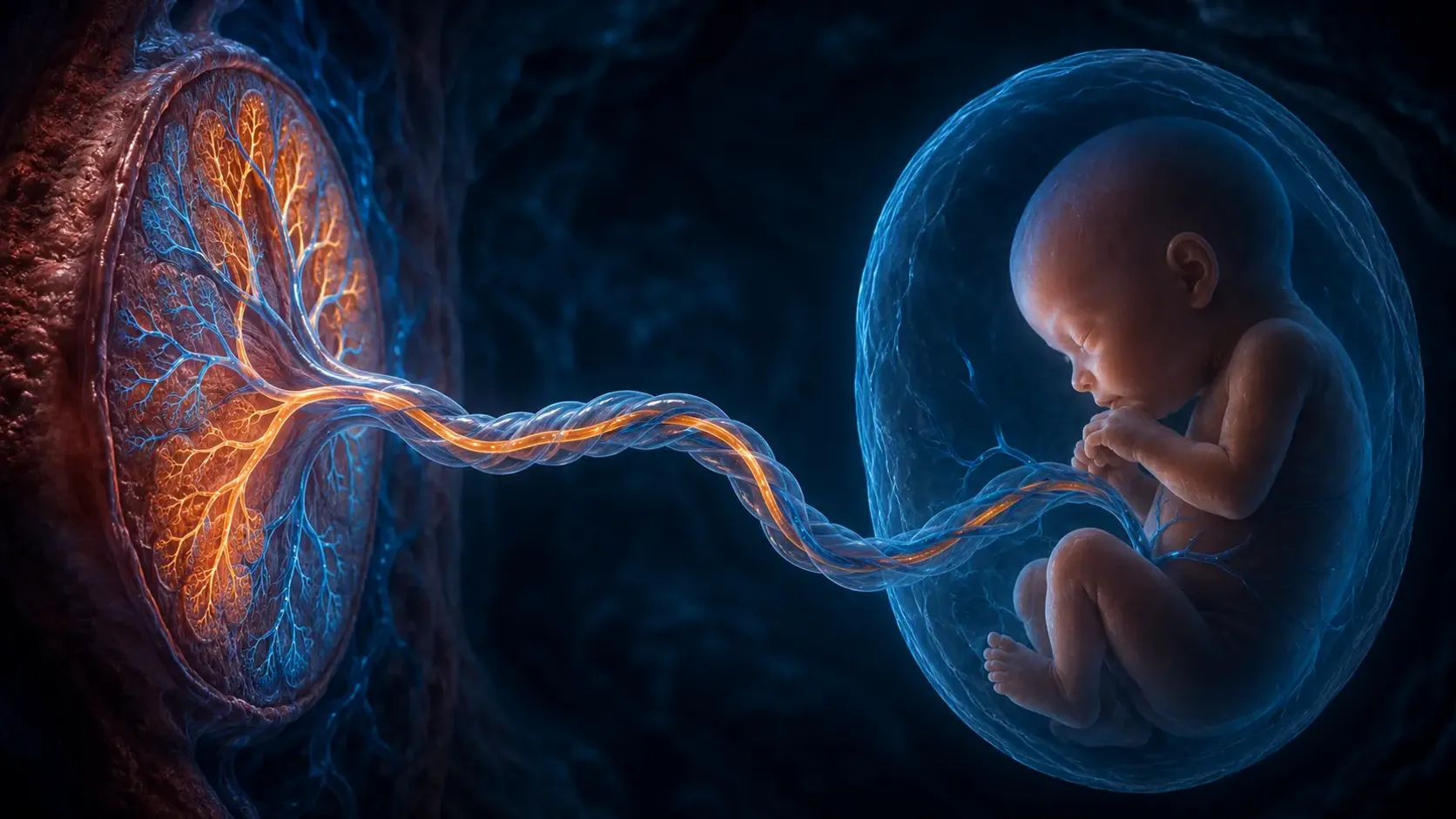
Maternal blood delivers oxygen to spaces surrounding microscopic placental villi. Fetal blood flows through capillaries inside those villi. The two supplies normally do not pour into one shared pool; gases and other molecules cross a thin interface between them. Oxygenated fetal blood then returns through the umbilical vein and is strategically streamed through the body.
The placenta is the gas-exchange organ before birth
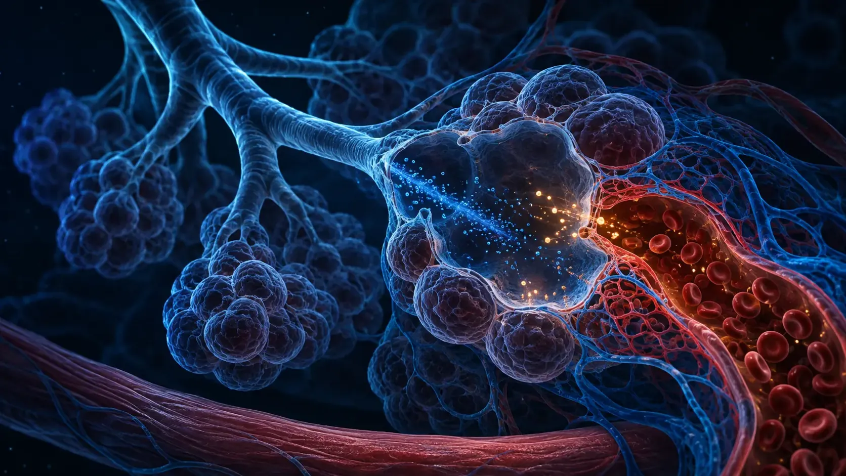
Maternal arteries deliver blood into the intervillous space of the placenta. Tree-like fetal villi project into that space, placing fetal capillaries close to the maternal supply across a thin tissue barrier. The arrangement creates a large surface for exchange without requiring normal direct mixing of maternal and fetal blood.
Oxygen moves down a partial-pressure gradient from maternal blood toward fetal blood. Carbon dioxide moves in the opposite direction and is removed by the mother's lungs. Nutrients, wastes and many signaling molecules also cross by mechanisms suited to their size and chemistry, which is why the placenta is much more than a passive filter.
The placenta itself consumes oxygen, and the efficiency of transfer depends on blood flow, surface area, barrier thickness and hemoglobin. Household observations cannot measure that exchange. Concerns about fetal movement or pregnancy health belong with maternity care rather than home attempts to infer oxygenation.
One umbilical vein carries oxygen toward the fetus
Names in the umbilical cord follow direction, not oxygen content. The single umbilical vein travels from the placenta toward the fetus and carries the most oxygenated blood in the fetal circulation. Two umbilical arteries carry lower-oxygen blood from the fetus back to the placenta for renewed exchange.
After entering the fetus, some umbilical venous blood supplies the liver. A substantial portion is directed through the ductus venosus toward the inferior vena cava and the right atrium. It mixes with returning blood, so fetal oxygen saturation is lower than the value typical of healthy adult arterial blood.
Lower saturation does not mean the healthy fetus is simply suffocating. Fetal hemoglobin has a higher oxygen affinity than adult hemoglobin, fetal blood has substantial oxygen-carrying capacity, and circulation patterns preferentially direct better-oxygenated streams toward organs with high developmental demand.
Temporary shunts let most blood bypass the fluid-filled lungs
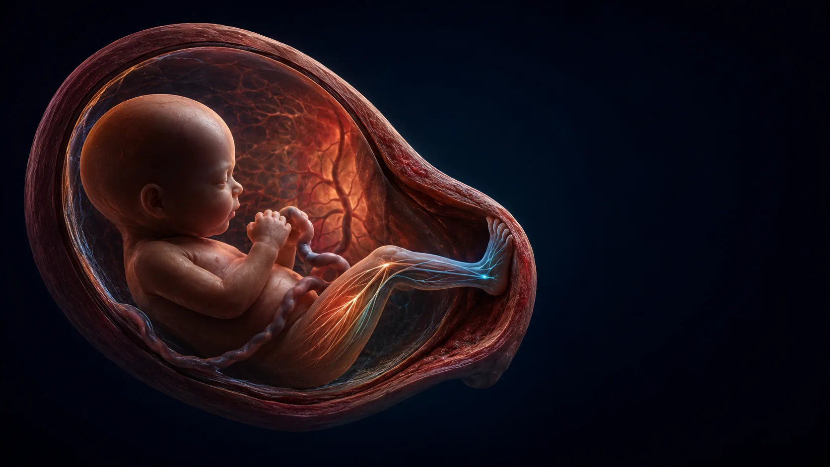
Blood reaching the right atrium can cross the foramen ovale into the left atrium, then move through the left ventricle and aorta. This route favors delivery of relatively well-oxygenated blood to the coronary circulation and brain. It is a normal fetal pathway, not a hole caused by disease.
Blood pumped from the right ventricle enters the pulmonary artery, but resistance in the unexpanded fetal lungs is high. Much of that flow therefore crosses the ductus arteriosus into the aorta instead of passing through the pulmonary capillaries. Only enough flow reaches the lungs to support their developing tissue.
The fetal heart consequently works with parallel pathways rather than the adult series in which the right heart sends blood through the lungs and the left heart sends oxygenated blood through the body. The temporary design matches a world where the placenta, not the lungs, is the exchange organ.
The first breaths trigger a rapid circulatory handoff
At birth, air enters the lungs, fluid is cleared and pulmonary vessels open as resistance falls. More blood now flows through lung capillaries and returns to the left atrium. Pressure relationships across the heart change, encouraging the flap of the foramen ovale to close functionally.
Separation from placental circulation removes the low-resistance placental pathway. Rising oxygen and changing chemical signals promote closure of the ductus arteriosus, while the ductus venosus and umbilical vessels also close. Anatomical sealing continues over different timescales after the first functional changes.
The newborn has not begun using a system that was previously idle in every sense—the lungs developed, produced fluid and received some blood before birth. But the source of gas exchange changes dramatically: the placenta is disconnected, the lungs inflate and the circulation is rebuilt around breathing air.
From maternal oxygen to fetal tissue
Maternal blood brings oxygen to the intervillous space. Oxygen crosses the placental interface into fetal capillaries and enters the umbilical vein, where fetal hemoglobin helps carry it at relatively low oxygen pressure.
The ductus venosus, foramen ovale and ductus arteriosus organize flow around the liver and lungs. After tissues use oxygen, the umbilical arteries return lower-oxygen blood to the placenta and the cycle begins again.
NCBI's fetal-circulation reference follows oxygen-rich blood from the placenta through the umbilical vein and fetal shunts.
A placental-flow review describes oxygen and nutrient transfer across thin membranes surrounding fetal villi.
Why it matters
Fetal circulation explains why an umbilical vein carries oxygenated blood while umbilical arteries carry lower-oxygen blood—the reverse of the pattern people often associate with veins and arteries.
It also makes birth easier to understand as a physiological transition. The first breaths, cord separation and vessel responses transform how the heart and lungs are connected within minutes.
Before birth, the placenta breathes on behalf of the fetus.
Oxygen crosses into fetal blood, returns through the umbilical vein and follows temporary shunts that largely bypass the fluid-filled lungs.
Research behind this story
We link to the primary study or an authoritative indexed review wherever possible. Caveats in the text reflect the limits of that evidence.
01