Quick answer
Your lungs work by coupling ventilation, diffusion and perfusion. The diaphragm and chest muscles enlarge the thoracic cavity, lowering pressure so air flows through the airways to alveoli. Across the extremely thin alveolar-capillary barrier, oxygen diffuses into blood and carbon dioxide diffuses into alveolar air. Hemoglobin carries most oxygen away, while pulmonary blood flow brings more carbon dioxide for removal. Surfactant reduces surface tension so alveoli are easier to keep open. Brainstem control adjusts breathing using chemical information, especially carbon dioxide and acidity, while local airway and vessel responses help match airflow with blood flow.
A breath feels like one action, but it solves three separate problems. Air must move into the chest, reach microscopic exchange surfaces and meet blood arriving at the right place and time. Failure at any one stage can reduce oxygen delivery even when the other stages still work.
The lungs achieve this through branching geometry. Conducting airways distribute air into smaller passages, alveoli place air beside an enormous capillary network, and the heart continually moves blood through that network. Pressure moves the breath; concentration gradients move the gases.
The diaphragm changes pressure rather than pulling on air
During a quiet inhalation, the diaphragm contracts and descends while external intercostal muscles help expand the rib cage. Thoracic volume increases, pressure inside the lungs falls slightly below atmospheric pressure and air flows inward. The lungs follow the chest wall because the pleural surfaces are mechanically coupled across a thin fluid layer.
Quiet exhalation is usually passive. Inspiratory muscles relax, elastic lung tissue recoils and air moves out as pressure rises. Exercise, coughing or breathing difficulty can recruit abdominal and other accessory muscles to change pressure more forcefully.
This mechanical step is ventilation: moving air, not yet moving oxygen into tissue. Air in the conducting zone—from the nose through much of the bronchial tree—does not directly exchange gases. It is warmed, humidified and transported toward the respiratory zone.
Branching airways deliver air to a vast microscopic surface
The trachea divides into bronchi, which divide repeatedly into smaller bronchioles. Cartilage supports larger airways, while smooth muscle and elastic tissue become increasingly important in smaller passages. Mucus and cilia help trap and move particles away from delicate exchange regions.
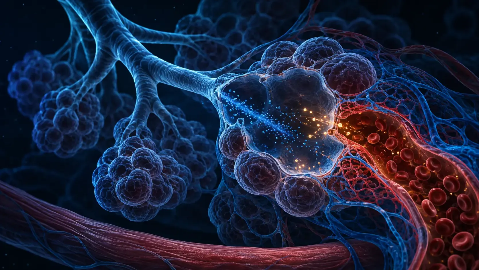
At the ends of respiratory bronchioles and alveolar ducts sit clusters of alveoli. Type I alveolar cells form most of the thin exchange surface. Type II cells produce surfactant, a mixture that lowers surface tension at the air-liquid interface and makes alveoli easier to inflate and stabilize.
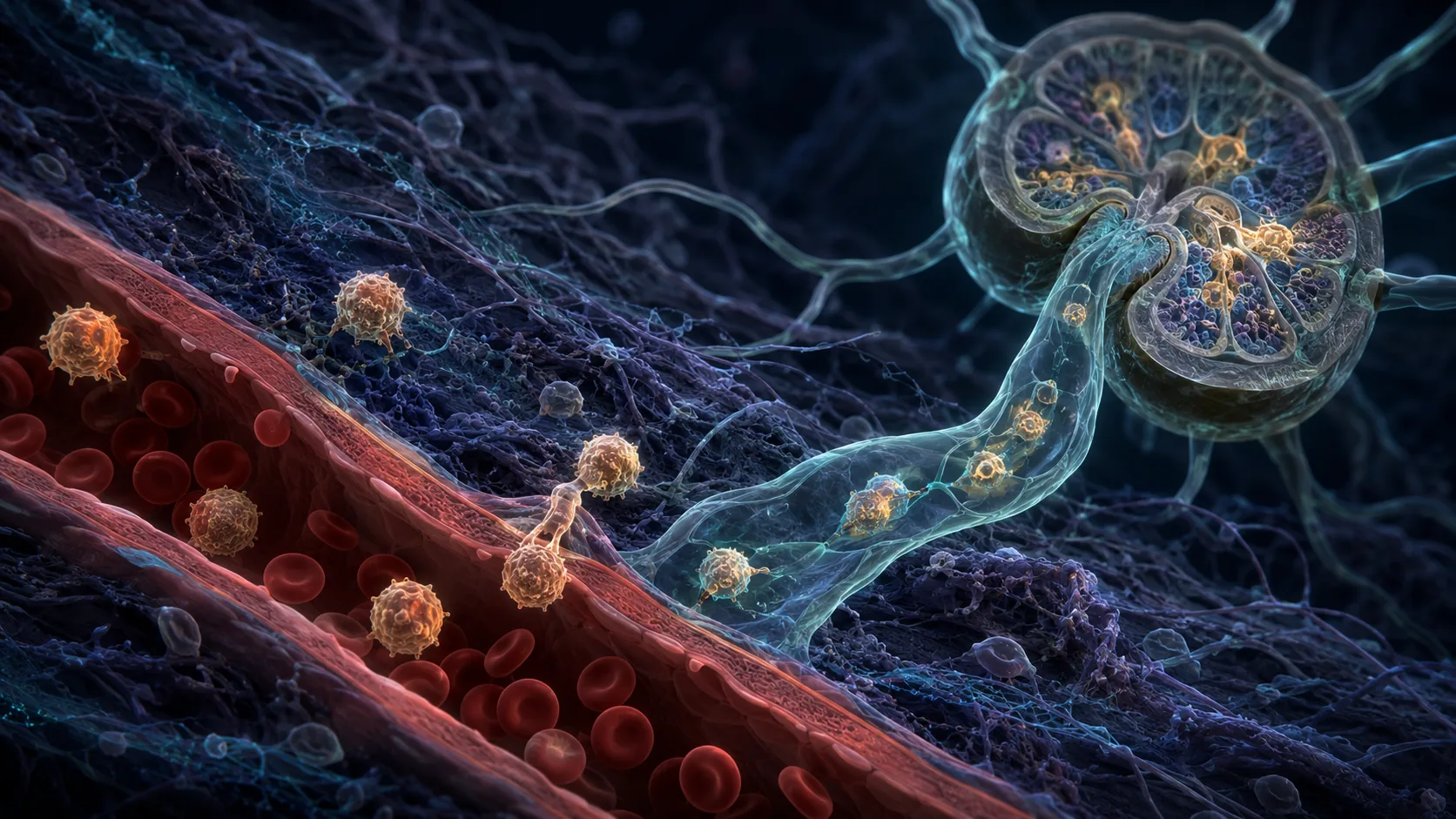
Alveolar macrophages patrol the surface for particles and microbes. Defense must remain measured: aggressive inflammation can thicken or flood the same barrier that gases need to cross, showing why protection and exchange have to coexist in very little space.
Oxygen and carbon dioxide diffuse in opposite directions
Fresh alveolar air has a higher oxygen partial pressure than the venous blood entering pulmonary capillaries. Oxygen therefore diffuses through surfactant, alveolar epithelium, shared basement structures and capillary endothelium into blood. Most then binds reversibly to hemoglobin inside red blood cells.
Carbon dioxide arrives in several chemical forms, much of it carried as bicarbonate. In the lungs those reactions reverse, and carbon dioxide diffuses from blood into alveoli before exhalation. Carbon dioxide crosses membranes readily, but its removal still depends on adequate ventilation.
Diffusion improves with greater surface area and a thinner barrier. Fluid, inflammation, scarring or lost alveolar surface can interfere. A person can move air yet exchange gases poorly, which is why breathing effort alone does not reveal blood oxygen or carbon dioxide precisely.
Airflow must meet blood flow, and breathing must match demand
The right side of the heart pumps blood through pulmonary arteries into capillaries surrounding alveoli. Effective exchange requires ventilation and perfusion to overlap. An alveolus receiving air but little blood contributes to dead space; blood passing poorly ventilated regions leaves without fully equilibrating.
Local responses help with matching. Low oxygen within a small lung region can constrict nearby pulmonary vessels, redirecting some blood toward better-ventilated alveoli. The system is imperfect, and widespread low oxygen can create a very different hemodynamic problem.
Breathing rhythm is generated in brainstem networks and adjusted by feedback. Carbon dioxide and the acidity it influences are powerful normal drivers; oxygen also matters, especially when levels fall. Exercise raises carbon dioxide production and oxygen demand, so ventilation and circulation increase together before conscious thought is required.
From a breath to oxygen delivery
Respiratory muscles expand the chest and air flows down branching airways. In alveoli, oxygen diffuses into capillary blood while carbon dioxide moves toward the airspace.
Hemoglobin carries oxygen through the body, and the heart returns carbon-dioxide-rich blood to the lungs. Brainstem and chemical feedback adjust the next breaths to keep the exchange aligned with metabolism.
The NHLBI explains how air reaches alveoli and oxygen enters blood while carbon dioxide leaves.
Pulmonary physiology references describe diffusion across the alveolar-capillary membrane and ventilation-perfusion matching.
Try it yourself
Observe a quiet breath without forcing it.
- Sit comfortably and place one hand lightly over the lower ribs.
- Notice the ribs and abdomen move as the diaphragm descends during a normal inhalation.
- Exhale without pushing and feel elastic recoil return the chest toward rest.
Do not perform prolonged breath-holding or forceful breathing. New or severe shortness of breath, blue or gray color, confusion or chest pain needs urgent medical assessment.
Why it matters
Separating ventilation, diffusion and perfusion explains why lung problems can look similar while arising through different mechanisms. Moving air is essential, but it is only one part of respiratory function.
The lungs also reveal a recurring biological tradeoff: the exchange surface must be extremely thin, open and exposed, yet it must defend itself continuously against particles and pathogens.
Your lungs work by bringing air and blood within a microscopic distance.
Muscles ventilate alveoli, gases diffuse across a thin barrier, and circulation carries them between the lungs and every tissue.
Research behind this story
We link to the primary study or an authoritative indexed review wherever possible. Caveats in the text reflect the limits of that evidence.
01