Quick answer
Babies kick in the womb because their developing nervous systems activate growing muscles and joints. Early movements are largely spontaneous motor patterns generated by the spinal cord and brain; with maturation they become more coordinated and responsive to sleep state, position and stimulation. Movement provides mechanical input that contributes to normal muscle, joint and bone development and helps rehearse patterns needed after birth. A pregnant person usually begins feeling movement during the second trimester, but timing and patterns vary. A noticeable reduction from an established movement pattern should be discussed promptly with a prenatal-care professional.
The first movements a pregnant person feels are often described as bubbles, fluttering or a tiny fish turning. Weeks later, the same hidden activity can become a clear kick that shifts the surface of the abdomen.
Movement starts long before that first sensation. As spinal circuits, brain networks, muscles and joints develop, the fetus produces an expanding repertoire of stretches, turns, startles, breathing-like motions and limb movements. A kick is one part of a developing motor system, not a deliberate message with one fixed meaning.
Movement begins before anyone can feel it
Ultrasound can detect embryonic and fetal movement early in development, before the fetus is large or strong enough for motion to be felt through the uterus and abdominal wall. The first perceived movement is called quickening and is commonly noticed somewhere in the middle of pregnancy, with substantial individual variation.
Perception depends on more than fetal activity. Fetal position, placental location, the thickness and sensitivity of surrounding tissues, previous pregnancy experience and attention can all influence when a movement becomes recognizable. An anterior placenta, for example, may cushion some sensations without stopping the fetus from moving.
The sensation changes as the fetus grows. Early flutters become taps, rolls, stretches and stronger pushes. Near the end of pregnancy the available space changes the character of movement, but healthy fetuses do not simply stop moving because the womb feels crowded.
The nervous system is building movement patterns
Motor neurons connect the spinal cord to skeletal muscle. Even before conscious control exists, developing neural circuits can generate patterned activity. Sensory feedback from moving joints and stretched tissues then returns information to the nervous system.
As the brain and spinal cord mature, movements become smoother, more varied and better coordinated. Fetal sleep and activity states also develop, so movement comes in episodes rather than at a constant rate. Quiet intervals can reflect normal sleep, while active periods may include whole-body motion.
A kick therefore does not require the fetus to decide to signal. It can emerge from spontaneous neural activity, reflexes and changing behavioral states. Responses to sound or maternal movement can occur later in gestation, but one kick cannot reveal a precise emotion or personality trait.
Why movement matters to muscles, joints and bones
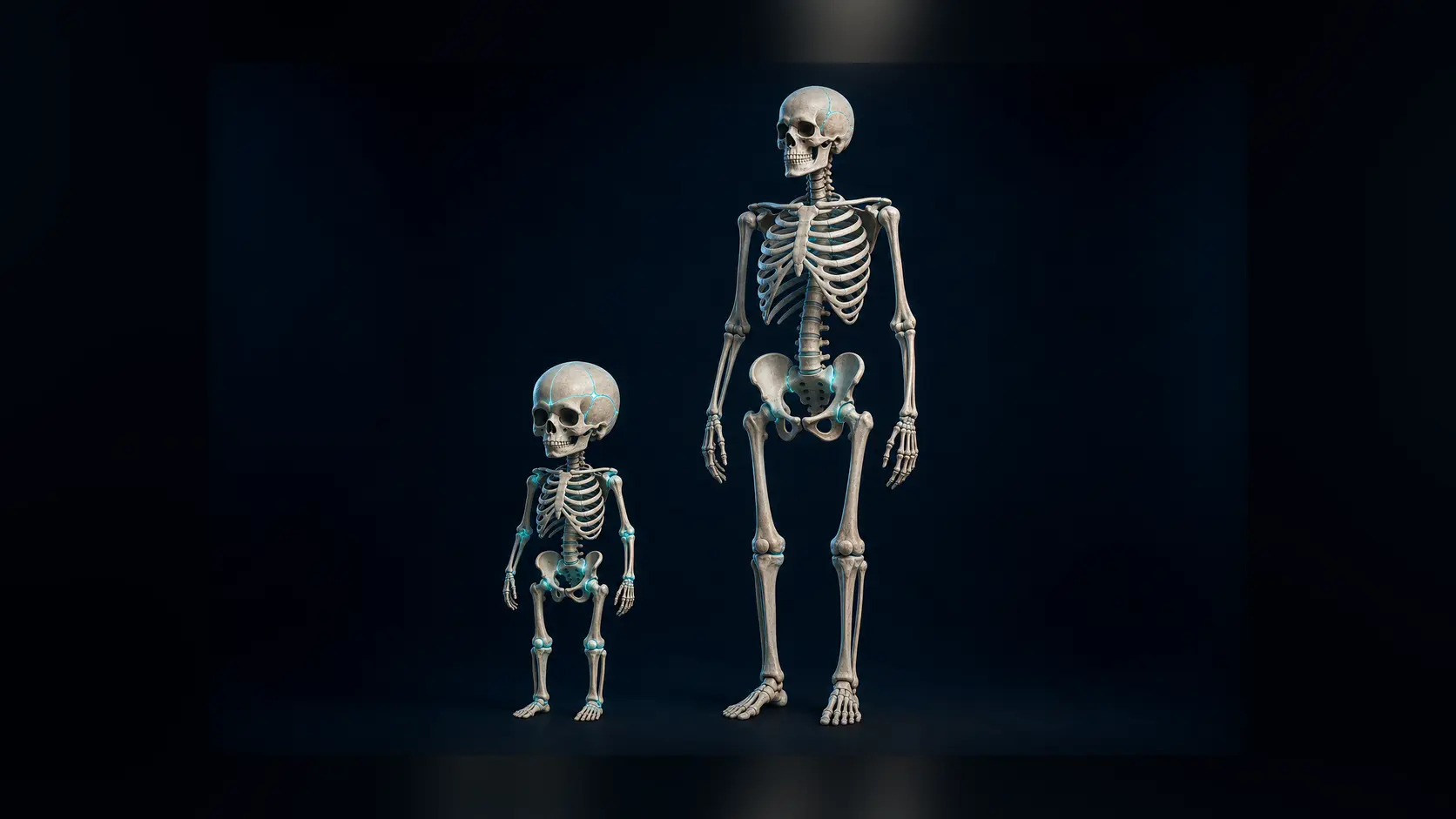
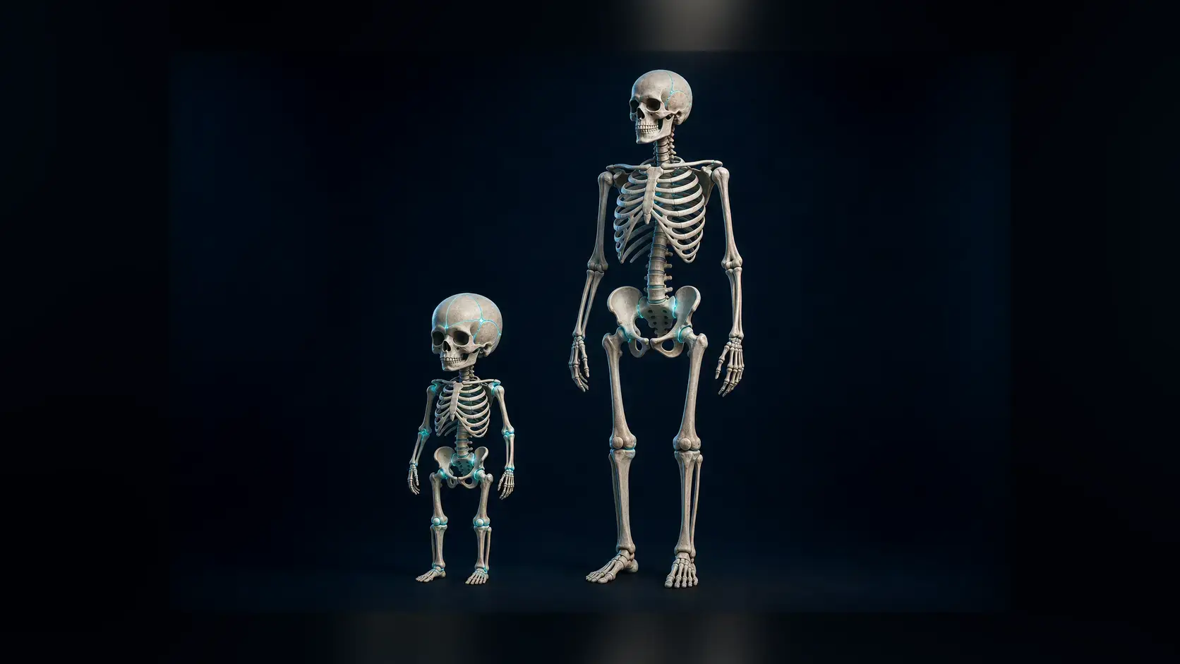
Contracting muscle loads tendons, joints and developing bone. Those mechanical forces help tissues organize and grow. Research on fetal biomechanics shows that reduced movement can alter joint shape and skeletal development, demonstrating that motion is part of normal construction rather than an optional extra.
Kicking also rehearses coordination. The womb supports the fetus in fluid, reducing weight-bearing while still allowing limbs to flex, extend and contact the uterine wall. These repeated cycles help prepare motor systems for breathing, feeding, posture and movement after birth.
This does not mean a stronger individual kick proves stronger bones or better health. Clinicians interpret movement as a pattern alongside gestational age and other assessments, not as a contest in intensity.
Why movement patterns matter more than a universal number
Fetuses differ in their usual timing and style of activity. Maternal meals, posture or time of day can change how movement is perceived, but no household trick can prove that fetal oxygenation or placental function is normal.
Prenatal teams may recommend awareness of the baby's established pattern or a structured counting method depending on clinical guidance. The important safety signal is a clear reduction or change from what is usual for that pregnancy—not comparing one person's count with someone else's online.
If movement is noticeably reduced or absent after it has become established, contacting the maternity or prenatal-care team promptly is safer than waiting for an article, app or home test to provide reassurance. Individual advice depends on gestational age and medical context.
From neural signal to felt kick
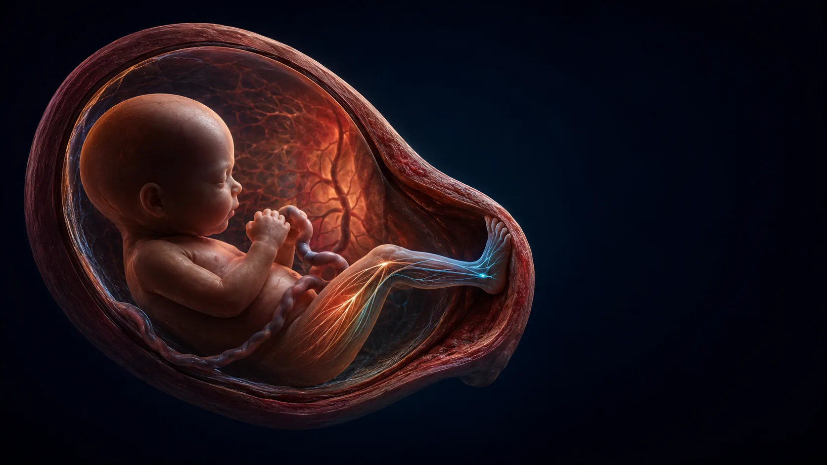
Developing motor circuits activate motor neurons, muscle fibres contract and a limb accelerates through amniotic fluid. When the foot or leg presses the uterine wall, force passes through the uterus and abdominal tissues.
If the movement is strong, close and well-positioned enough, sensory receptors in maternal tissue register it. The same movement may be obvious on ultrasound yet unfelt, or felt strongly from a different position.
A review explains how fetal movement can reflect neurological integrity and fetal wellbeing.
Biomechanical studies model how lower-limb movement loads the uterine environment.
Try it yourself
Learn the usual pattern without provoking movement.
- Notice when movement is normally active or quiet over several days.
- Use a counting method only if your prenatal-care team recommends one and explains it.
- Report a noticeable reduction rather than relying on cold drinks, sound or repeated poking for reassurance.
Reduced fetal movement can need prompt assessment. Contact the appropriate maternity or prenatal service for individual advice.
Why it matters
Fetal movement makes development tangible: nervous-system activity becomes muscle force, and muscle force helps shape the skeleton that movement will later control.
It also carries clinical information, which is why a meaningful change in an established pattern should be taken seriously rather than interpreted through social-media rules.
A kick is development in motion.
Neural circuits activate growing muscles, movement loads developing joints and bones, and the resulting force can travel through the uterine wall to be felt.
Research behind this story
We link to the primary study or an authoritative indexed review wherever possible. Caveats in the text reflect the limits of that evidence.
01