Quick answer
Growth plates lengthen many childhood bones through endochondral ossification. Resting chondrocytes supply cells that enter organized columns, divide and then enlarge. Cartilage matrix around the enlarged cells is prepared for mineralization, blood vessels and bone-forming cells advance from the shaft side, and the cartilage scaffold is replaced by bone. Repeating this sequence adds new material near each end while the existing shaft remains rigid. During puberty, sex-steroid signaling first supports the growth spurt and later accelerates growth-plate maturation and closure, ending further lengthening.
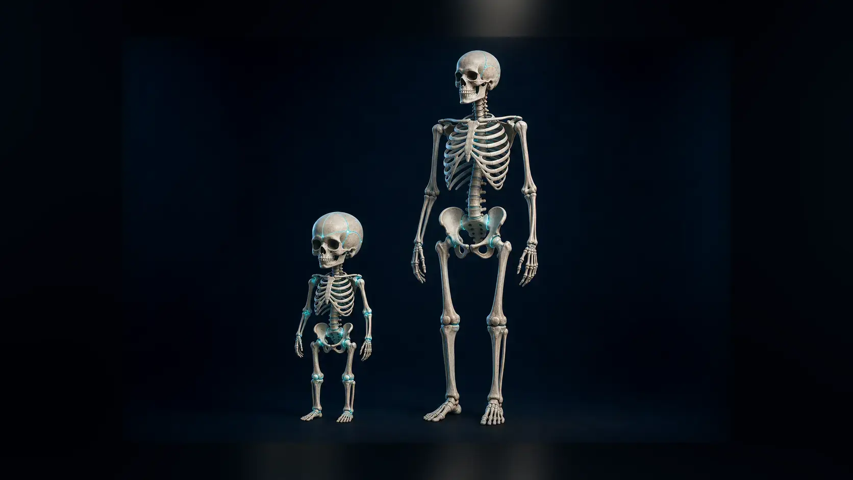
A femur can become much longer between infancy and adulthood, yet its hard shaft cannot simply stretch. Growth happens in a narrow band of cartilage near each end of the bone, where cells create new space before bone tissue takes over.
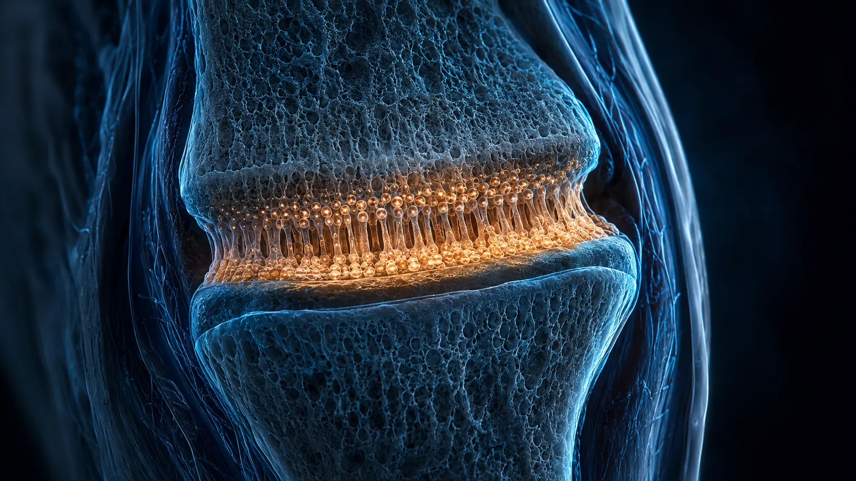
Those bands are growth plates, or physes. Their microscopic order turns cell division, swelling and mineralization into centimeters of height while preserving joints at the ends.
A growth plate is an organized cellular factory
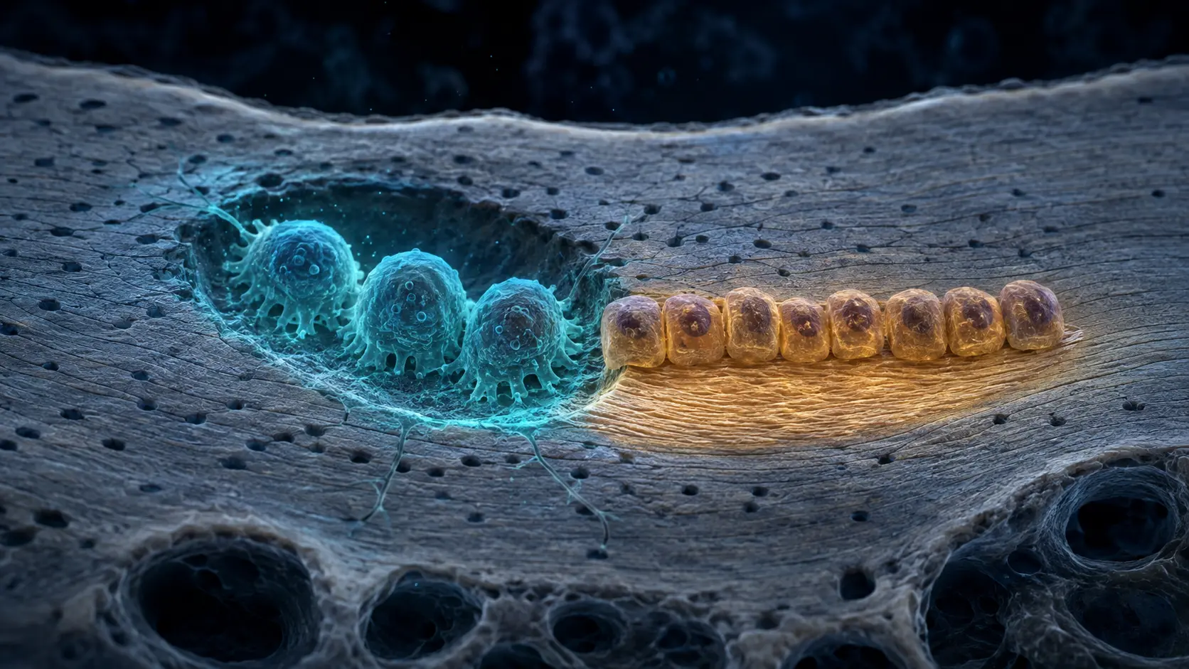
The plate sits between the epiphysis at the joint end and the metaphysis toward the shaft. It is not one uniform strip. Researchers describe resting, proliferative and hypertrophic zones, followed by regions where cartilage calcifies and new bone is deposited.
In the proliferative zone, chondrocytes divide and align in columns parallel to the bone's long axis. Farther along, hypertrophic chondrocytes expand dramatically and alter the surrounding matrix. Cell enlargement is a major contributor to the rate of longitudinal growth, not merely a prelude to it.
Capillaries, osteoblast-lineage cells and other marrow elements approach from the metaphyseal side. Mineralized cartilage provides a temporary scaffold on which new bone is laid. The result is a moving construction front rather than a fixed seam being pulled apart.
Local programs and whole-body hormones set the pace
Growth hormone and insulin-like growth factor 1 influence chondrocyte proliferation and maturation, but they are part of a larger network. Thyroid hormone, glucocorticoids, nutrition, oxygen conditions and local signals such as Indian hedgehog, PTH-related protein and FGFR3 all affect growth-plate behavior.
That complexity explains why height cannot be reduced to one hormone or one food. Genetics influences the potential trajectory, while health, nutrition, chronic disease and endocrine conditions can modify how closely growth follows it.
Mechanical loading also interacts with the plate. Normal movement belongs to healthy development, but a growth plate is mechanically vulnerable because it is cartilage between stronger bone regions. A fracture that crosses it may disturb later growth and needs proper clinical evaluation.
Puberty speeds growth before the plates close
Early in puberty, coordinated growth-hormone and sex-steroid changes help produce the adolescent growth spurt. Estrogen signaling is important in all sexes; in many males, some testosterone is converted to estrogen by aromatase.
With continued maturation, the supply and proliferative capacity of growth-plate cells decline. The cartilage zone narrows and is eventually replaced by bone, leaving an epiphyseal line. Once fusion is complete, that bone can remodel and thicken but cannot resume normal longitudinal growth through the former plate.
Closure occurs at different times in different bones and people, so a birthday cannot reveal whether a plate is open. Clinicians may use history, examination and appropriately interpreted imaging when growth or injury raises a genuine medical question.
How cartilage becomes added bone length
Chondrocytes divide in columns and then enlarge, pushing the epiphysis away from the shaft while maintaining the cartilage template.
On the shaft side, vessels and bone-forming cells replace that template with mineralized bone. Balanced repetition advances the end of the bone.
A physiological review describes the growth plate as the site where longitudinal bone growth is organized into distinct cartilage zones.
Why it matters
Growth plates connect microscopic cell behavior with visible childhood growth and the timing of puberty.
They are also clinically important structures: injuries, endocrine disorders and chronic illness can alter growth, while ordinary variation cannot be diagnosed from height alone.
Children grow taller by building new bone at cartilage plates.
Ordered columns of chondrocytes create length, and ossification converts the new cartilage scaffold into durable bone.
Research behind this story
We link to the primary study or an authoritative indexed review wherever possible. Caveats in the text reflect the limits of that evidence.
01