Quick answer
Yes—the liver can restore lost tissue after a partial surgical removal or some acute injuries. Most of this response comes from mature liver cells called hepatocytes re-entering the cell cycle and dividing, supported by signals from immune, endothelial and stellate cells. It is better described as compensatory growth than as rebuilding the exact missing lobe: the remaining lobes enlarge until adequate mass and function return. The result depends on how much healthy tissue remains, blood flow, age and disease. Chronic inflammation, fatty liver disease, fibrosis and cirrhosis can weaken or distort the regenerative response.
Remove part of a healthy liver and the organ can recover much of the mass it lost. That sounds like the regrowth of a salamander limb, but the geometry and cellular strategy are different. The cut-away lobe does not usually reappear as a miniature copy that grows into shape.
Instead, cells in the liver tissue that remains switch from routine metabolic work into a coordinated growth response. Existing lobes enlarge, blood vessels and supporting tissue adapt, and growth slows once the organ has recovered enough functional capacity for the body.
The liver restores mass, not the original silhouette
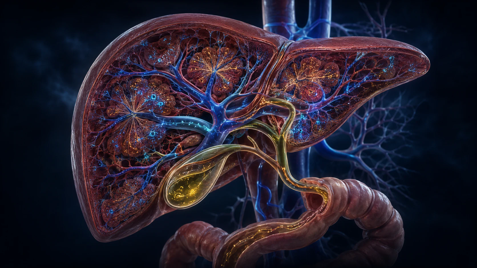
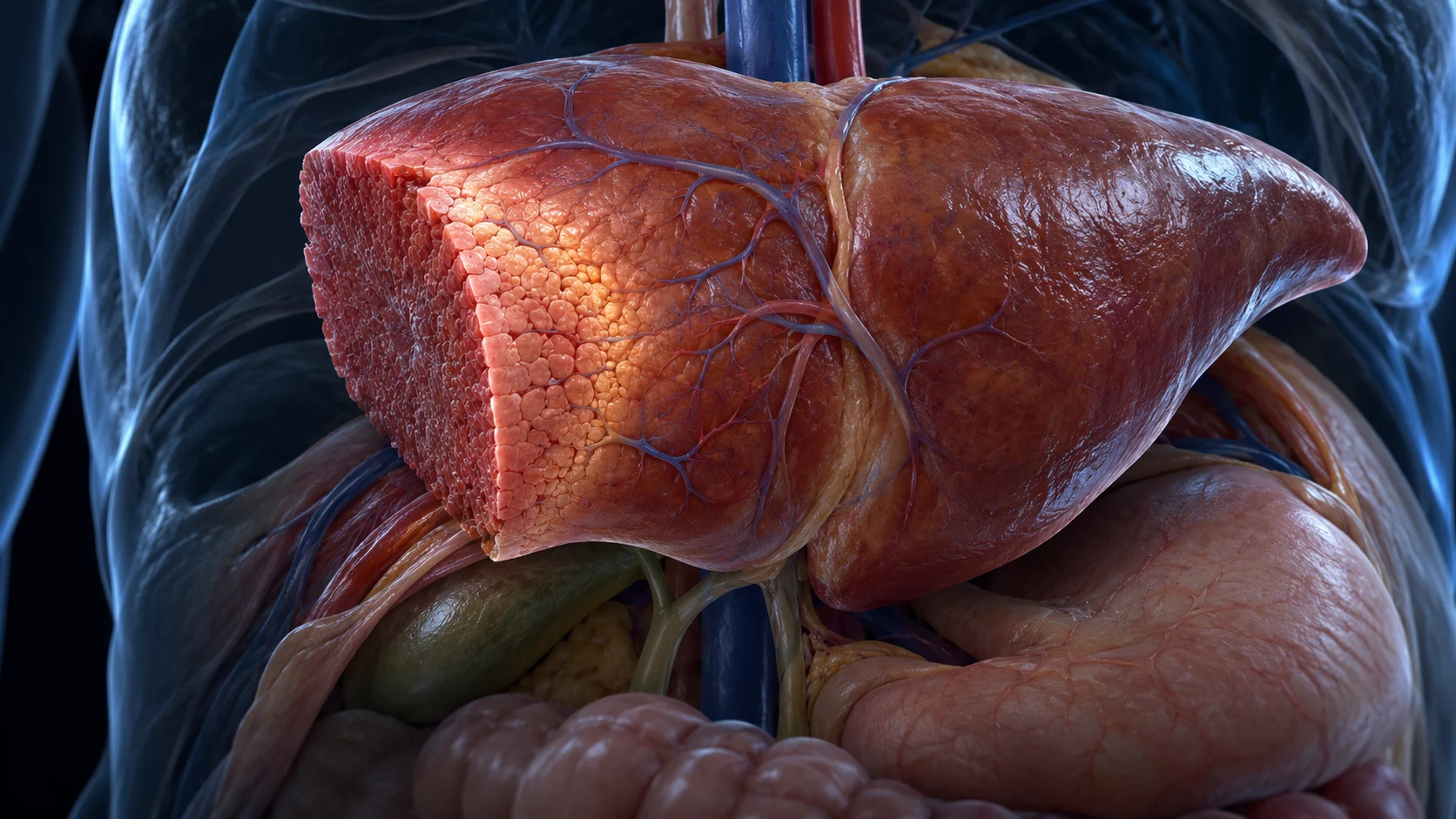
A classic way to study liver regeneration is partial hepatectomy, an operation that removes a portion of the organ without directly injuring the tissue left behind. The remaining lobes respond rapidly. Their cells grow and divide, and the lobes expand until liver mass is better matched to the body's needs.
That distinction matters. A regenerating limb must rebuild a map of joints, muscles, nerves, skin and digits. The liver mainly has to restore enough organized tissue to perform shared functions such as metabolism, protein synthesis and bile production. Its repeated microscopic units make compensatory growth possible without recreating the missing outline.
Even the word regenerate needs care. Researchers use it because lost functional tissue is restored, but the process combines cell proliferation, cell enlargement, vascular remodeling and changes in the extracellular matrix. It is not a perfect anatomical rewind.
Mature hepatocytes do most of the rebuilding
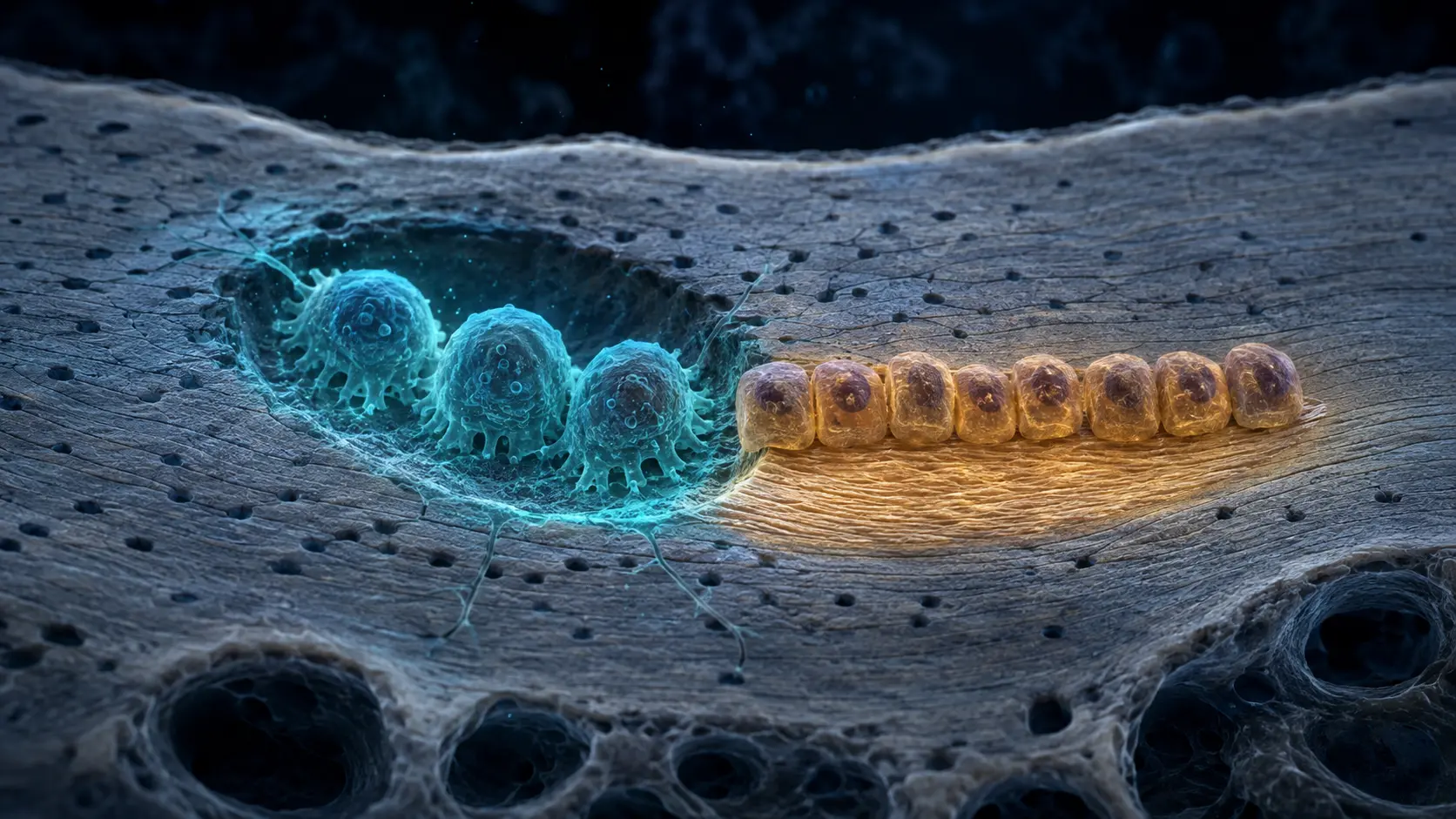
In an uninjured adult liver, most hepatocytes are not actively dividing. After tissue loss, inflammatory signals and growth factors help prime them to leave that quiet state. Hepatocyte growth factor, epidermal growth factor family signals, interleukin-6 and several other pathways help coordinate entry into the cell cycle.
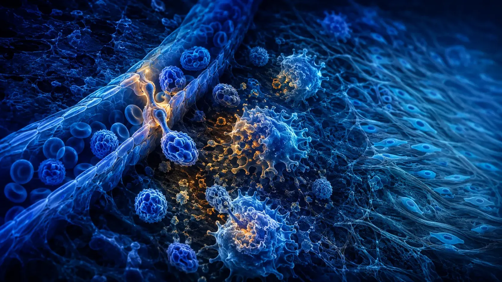
The response is a community effort. Kupffer cells, the liver's resident macrophages, release and interpret inflammatory signals. Sinusoidal endothelial cells line the specialized blood channels and help rebuild the vascular network. Stellate cells and other stromal populations reshape the matrix and exchange signals with hepatocytes.
When ordinary hepatocyte proliferation is severely impaired, other cell states may contribute under particular experimental or disease conditions. But current evidence does not support the simple story of one universal liver stem cell routinely rebuilding the organ. In common acute regeneration, proliferation of surviving hepatocytes is the central mechanism.
A change in blood flow helps announce the loss
The liver has an unusual circulation: portal blood from the digestive organs and arterial blood both enter it. After part of the liver is removed, the same incoming portal flow is directed through less tissue. Changes in pressure, stretch and molecules arriving from the gut help alter the local signaling environment.
Cells then balance two demanding jobs. The remnant must continue regulating nutrients, processing chemicals and making proteins while also producing new cellular material. Metabolism changes to support both survival and growth, and new or remodeled sinusoids must keep expanding tissue supplied.
No single trigger runs the whole program. Mechanical forces, cytokines, growth factors, bile-acid signaling, hormones and nutrients overlap. Their timing and location help explain why laboratory studies that activate one pathway rarely reproduce complete, controlled regeneration on their own.
Successful regeneration has to know when to stop
Growth that starts but never stops would be dangerous. As useful mass returns, pro-growth signals fade and inhibitory networks regain influence. Pathways involving transforming growth factor beta, Hippo signaling and metabolic feedback are among the systems studied in this termination phase.
The stopping rule is not a simple organ-size ruler. Researchers think the liver integrates blood flow, metabolic workload, cell contacts, matrix tension and signals from other organs. The endpoint is therefore a functional balance between liver mass and the needs of the body.
This control also connects regeneration research with cancer biology. Many pathways that help normal cells divide after injury can contribute to tumors if they remain active or acquire mutations. A safe regenerative response depends as much on restraint and tissue organization as on fast proliferation.
How well it grows back depends on the liver it starts with
There is no single countdown for a liver to grow back. After planned surgery, measurable volume can return over weeks and continue changing for months, but volume is not identical to microscopic organization or full function. The amount removed, surgical method, blood supply and the person's health all influence the course.
Acute tissue loss and chronic liver disease create different biological problems. Persistent inflammation, metabolic injury or viral damage repeatedly kills cells and promotes fibrotic scar. Over time, senescent cells, altered blood flow and dense matrix can make regeneration less efficient even while nodules of surviving cells grow.
That is why the liver's regenerative reputation should never be treated as permission to ignore alcohol-related injury, hepatitis, medication toxicity or metabolic disease. Regenerative capacity is substantial, not unlimited. Questions after liver surgery, donation or a diagnosis require advice from the treating clinical team, not a generic timeline.
From tissue loss to restored working mass
Tissue loss changes blood flow and releases injury-related signals. Surviving hepatocytes and supporting cells coordinate proliferation, matrix remodeling and vascular growth while the remnant keeps performing essential metabolic work.
As the liver regains enough capacity, growth-promoting signals decline and inhibitory feedback strengthens. The remaining lobes are larger, but the removed lobe has not been rebuilt as an identical structure.
A recent review describes how hepatocytes and neighboring cell types coordinate liver regeneration.
Research on chronic injury explains why persistent inflammation and fibrosis can exhaust or distort repair.
Why it matters
Liver regeneration makes partial liver donation and many liver resections possible, while also showing why organ function can return without exact anatomical replacement.
Its limits explain why acute injury cannot be used as a model for every chronic disease. A liver trapped in repeated damage and fibrosis faces a different problem from a healthy liver after planned surgery.
The liver can restore capacity without recreating the missing piece.
Surviving hepatocytes and supporting cells expand the remaining tissue, then feedback systems slow growth. Chronic disease and scarring can sharply reduce that ability.
Research behind this story
We link to the primary study or an authoritative indexed review wherever possible. Caveats in the text reflect the limits of that evidence.
01