Quick answer
Apoptosis is a regulated form of cell death in which caspase enzymes dismantle a cell. Internal stress can shift BCL-2-family proteins toward mitochondrial outer-membrane permeabilization, releasing cytochrome c and assembling an apoptosome that activates initiator caspase-9. External death receptors can activate caspase-8. Both routes engage executioner caspases that cut selected proteins, condense chromatin and help form membrane-bound apoptotic bodies. Phosphatidylserine exposed on the cell surface attracts phagocytes. Because contents remain contained and are cleared quickly, apoptosis usually provokes less inflammation than lytic death, but the boundary is not absolute.
A living tissue cannot preserve itself by keeping every cell forever. Some cells become unnecessary after development, accumulate irreparable damage or pose a threat to their neighbors. Removing them safely is as important as making replacements.
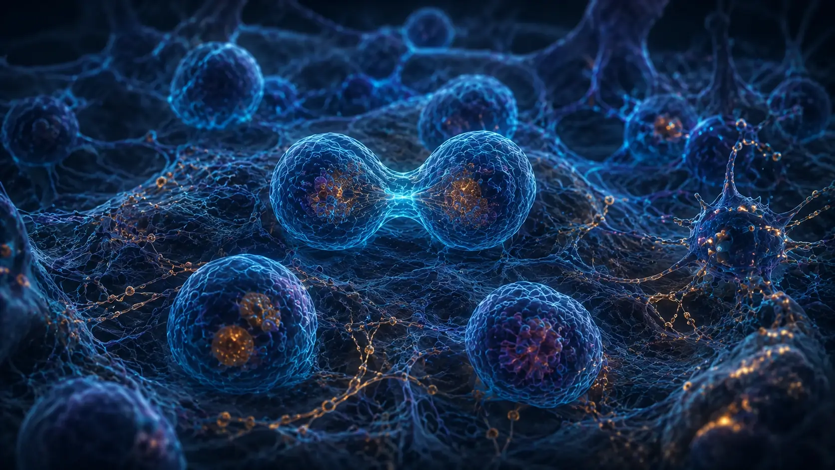
Apoptosis is one regulated solution. Instead of swelling until the membrane bursts, a cell activates a protease program, condenses its nucleus, shrinks and divides its contents into packages. Nearby cells recognize an 'eat me' signal and clear the remains.
The dying cell changes shape in a recognizable sequence
An apoptotic cell becomes smaller and detaches from some neighbors. Chromatin condenses against the nuclear envelope, DNA is fragmented in an organized pattern, and the plasma membrane forms blebs while initially preserving its barrier.
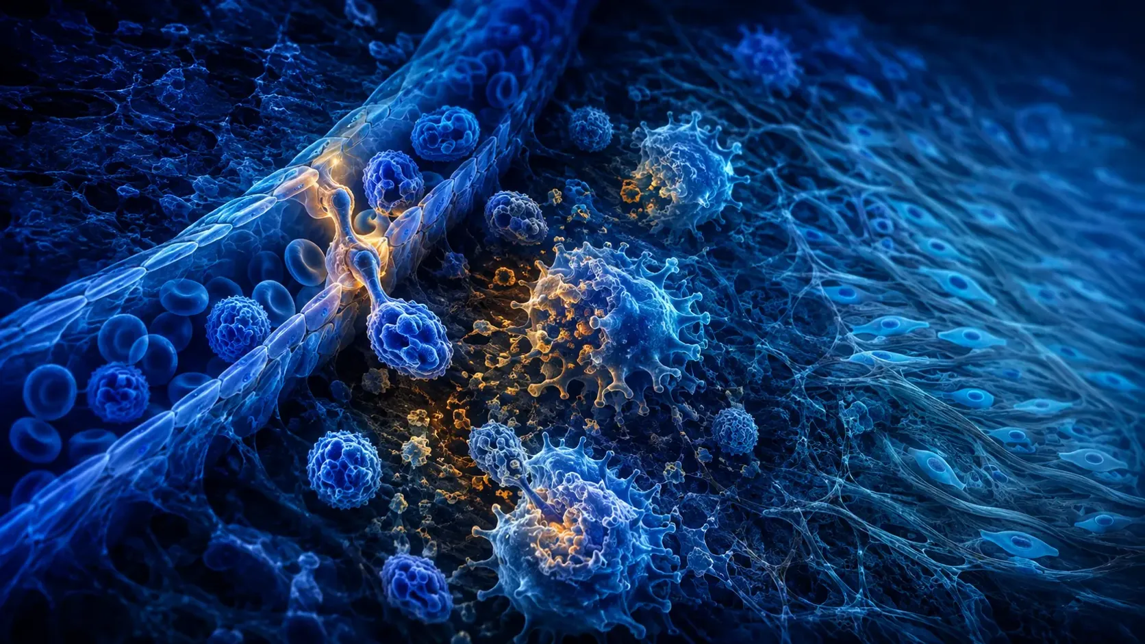
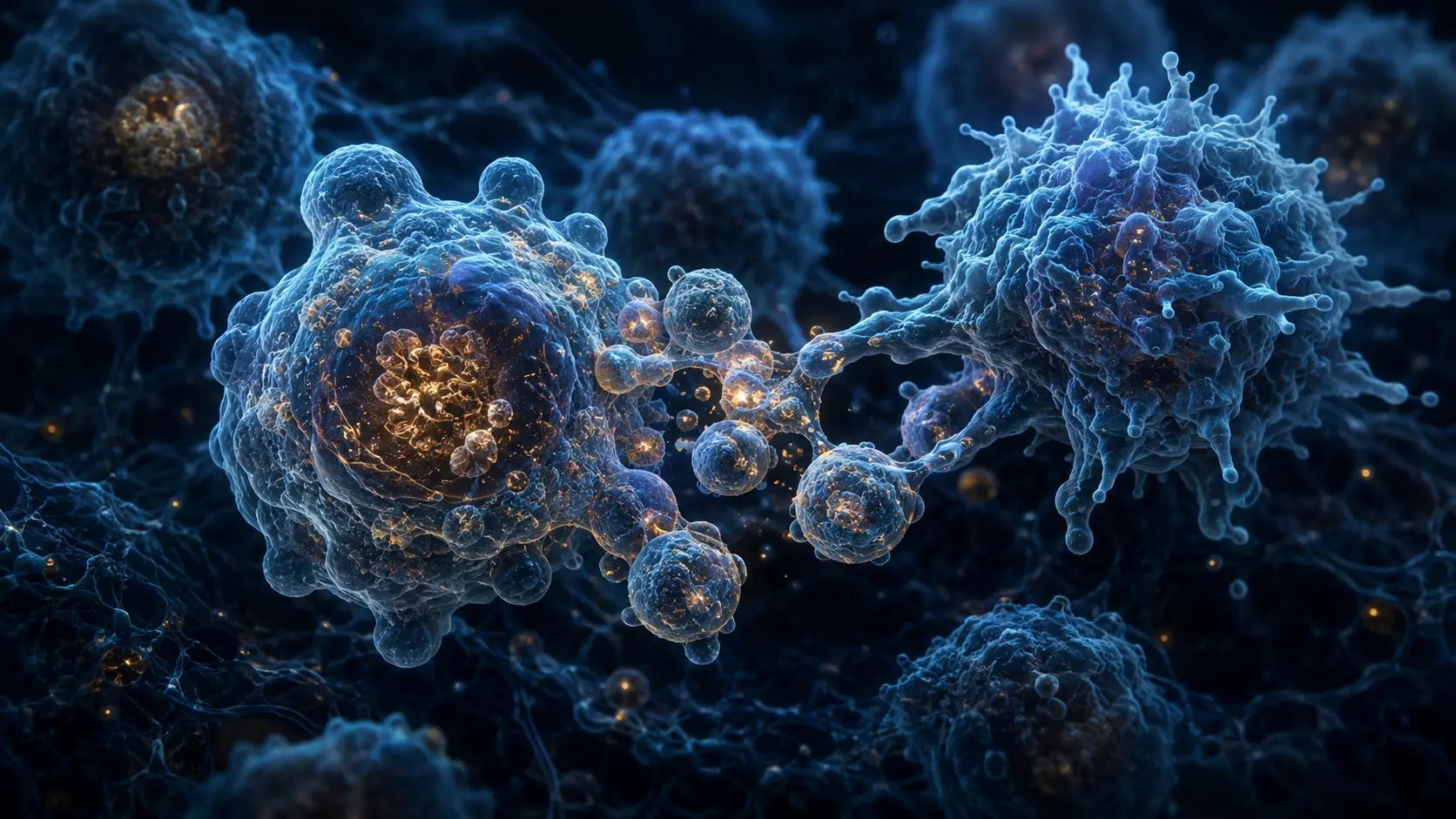
The cell separates into membrane-bound apoptotic bodies containing cytoplasm and organelle fragments. Macrophages or nearby cells engulf them. Efficient clearance prevents intracellular enzymes and damage signals from spreading widely through tissue.
These features are useful but not a perfect label from one image. Modern cell-death classification emphasizes molecular mechanisms because morphology can overlap and a cell may move from one death program into secondary membrane rupture when clearance fails.
The intrinsic pathway measures internal stress
DNA damage, growth-factor withdrawal, oncogene stress or severe organelle dysfunction can alter the balance of BCL-2-family proteins. Some members preserve mitochondrial integrity; others activate the pore-forming proteins BAX and BAK.
Once the mitochondrial outer membrane becomes permeable, cytochrome c enters the cytosol. With APAF1 and procaspase-9 it helps assemble the apoptosome. Activated caspase-9 then engages executioner caspases, especially caspase-3 and caspase-7.
This control is not one linear switch. Anti-apoptotic proteins, BH3-only sensors, cellular metabolism and tissue context shape the threshold. Mitochondrial permeabilization is often a point of no return, but cells and experiments do not all behave identically.
External receptors can launch a second route
Death receptors such as Fas and selected tumor-necrosis-factor receptor family members respond to extracellular ligands. Receptor complexes recruit adaptor proteins and procaspase-8, creating a platform that activates the enzyme.
Caspase-8 can directly activate executioner caspases or amplify the mitochondrial pathway through BID. This cross-talk lets immune cells remove infected, damaged or unwanted targets while linking the extrinsic and intrinsic programs.
Caspases are proteases with controlled substrates, not indiscriminate digestive fluid. They cleave structural and regulatory proteins in a coordinated sequence while nucleases fragment DNA and membrane changes prepare the cell for recognition.
Clearance determines how quiet the death remains
Healthy cell membranes keep phosphatidylserine mainly on the inner leaflet. During apoptosis it appears on the outer surface, where bridging molecules and phagocyte receptors recognize it. Engulfment can also promote anti-inflammatory signaling.
If apoptotic cells are not cleared, their membranes eventually lose integrity in secondary necrosis and release contents. Apoptosis is therefore usually immunologically quiet, not inherently invisible or always anti-inflammatory.
Too little apoptosis can allow damaged or autoreactive cells to persist; too much can contribute to tissue loss. Cancer, autoimmunity, infection and neurodegeneration can involve different failures of death control, but translating one pathway into treatment requires disease-specific evidence.
From a death signal to clean removal
Internal stress or a surface death receptor activates an initiator-caspase pathway. Mitochondrial signaling uses BCL-2-family control, cytochrome c and the apoptosome.
Executioner caspases dismantle selected structures. The cell exposes phosphatidylserine, fragments into contained bodies and is engulfed before its contents spill.
The Nomenclature Committee on Cell Death distinguishes apoptosis using mechanism-based criteria within regulated cell death.
Why it matters
Apoptosis explains how embryos sculpt structures, immune systems edit cell populations and adult tissues remove damaged cells without routinely inflaming their surroundings.
Its regulation also shows why cell survival is an active balance: blocking death can support cancer, while excessive death can destroy irreplaceable tissue.
Some cells protect the organism by leaving cleanly.
Caspases coordinate dismantling, membranes contain the fragments and phagocytes complete the process before dangerous contents spread.
Research behind this story
We link to the primary study or an authoritative indexed review wherever possible. Caveats in the text reflect the limits of that evidence.
01