Quick answer
Skin heals a cut through four overlapping processes. Hemostasis constricts vessels and forms a platelet–fibrin clot. Inflammation recruits immune cells to remove damaged material and coordinate repair. During proliferation, keratinocytes cover the surface, new capillaries grow, fibroblasts produce extracellular matrix and the wound contracts. Remodeling then reorganizes collagen for months. A scar forms when rapid closure replaces the original basket-like skin architecture with denser, more aligned collagen. It restores the barrier and much of the strength, but usually not hair follicles, sweat glands or the exact structure of uninjured skin.
Within seconds of a cut, damaged vessels constrict and platelets begin building a temporary seal. Long before the surface looks closed, cells beneath it are exchanging signals about debris, microbes, blood supply and mechanical tension.
Textbooks divide healing into phases, but the body does not wait for one phase to finish before starting the next. Hemostasis, inflammation, proliferation and remodeling overlap, and that overlap is one reason a scar can keep changing for months.
The first structure is a temporary seal
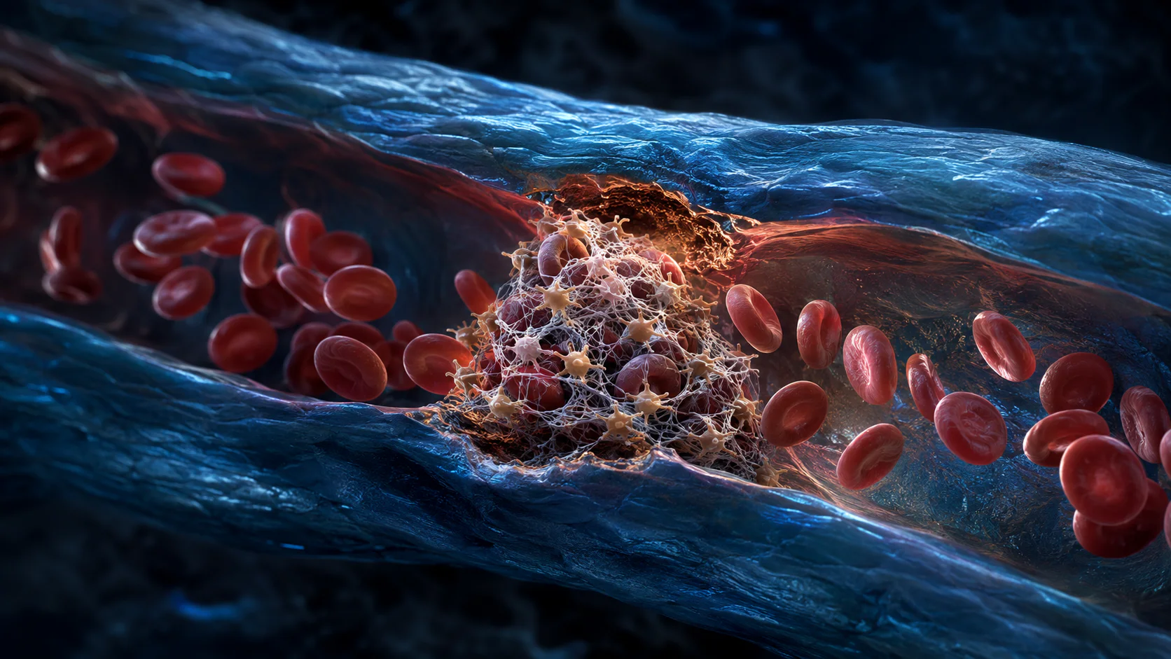
Platelets adhere to exposed material in the damaged vessel wall and activate one another. The clotting cascade generates fibrin strands that stabilize the platelet plug and trap blood cells in a provisional matrix.
That clot limits blood loss, but it also becomes a signaling platform. Platelets release molecules that recruit immune cells and influence fibroblasts, endothelial cells and surface keratinocytes.
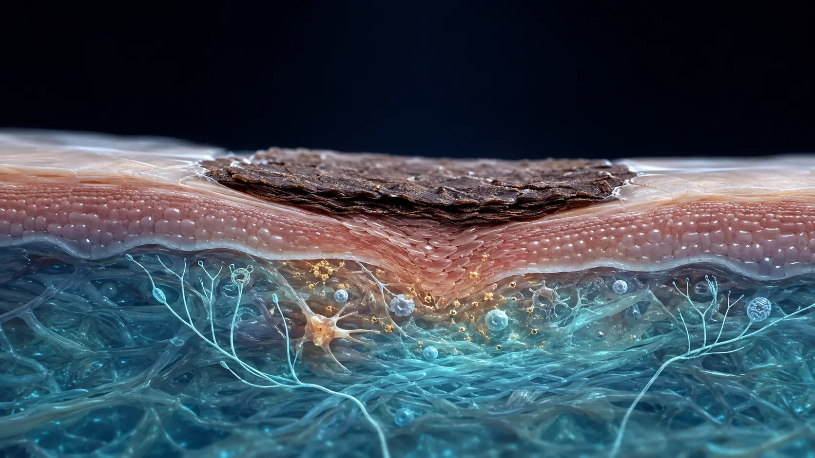
A dry scab may form above the clot when blood and tissue fluid meet air. The biologically active repair field extends deeper than the visible crust.
Inflammation clears damage and changes the program
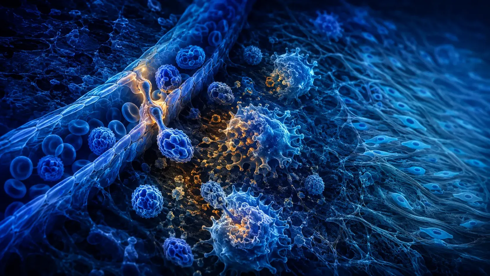
Neutrophils arrive early and help control contamination. Monocytes enter tissue and become macrophages that engulf debris, coordinate inflammation and later promote tissue construction and resolution.
Inflammation is therefore necessary, not simply a mistake to eliminate. Too little can leave contamination and debris; excessive or prolonged inflammation can delay closure and favor abnormal scarring.
The shift from an inflammatory to a repair-supporting environment is gradual. Researchers know many of the participating signals, but the complete timing network in human wounds remains an active field of study.
New tissue fills the gap while the surface closes
Keratinocytes migrate from the wound edges and surviving skin structures to restore the epidermal barrier. Endothelial cells build capillary loops, giving granulation tissue its red, vascular appearance.
Fibroblasts deposit collagen, fibronectin and other extracellular-matrix components. Some acquire contractile features and pull wound edges inward, reducing the area that must be rebuilt.
These events overlap with immune activity. Oxygen supply, infection, diabetes, nutrition, medicines and repeated mechanical stress can alter their speed, which is why a calendar alone cannot judge an individual wound.
A scar favors fast closure over perfect reconstruction
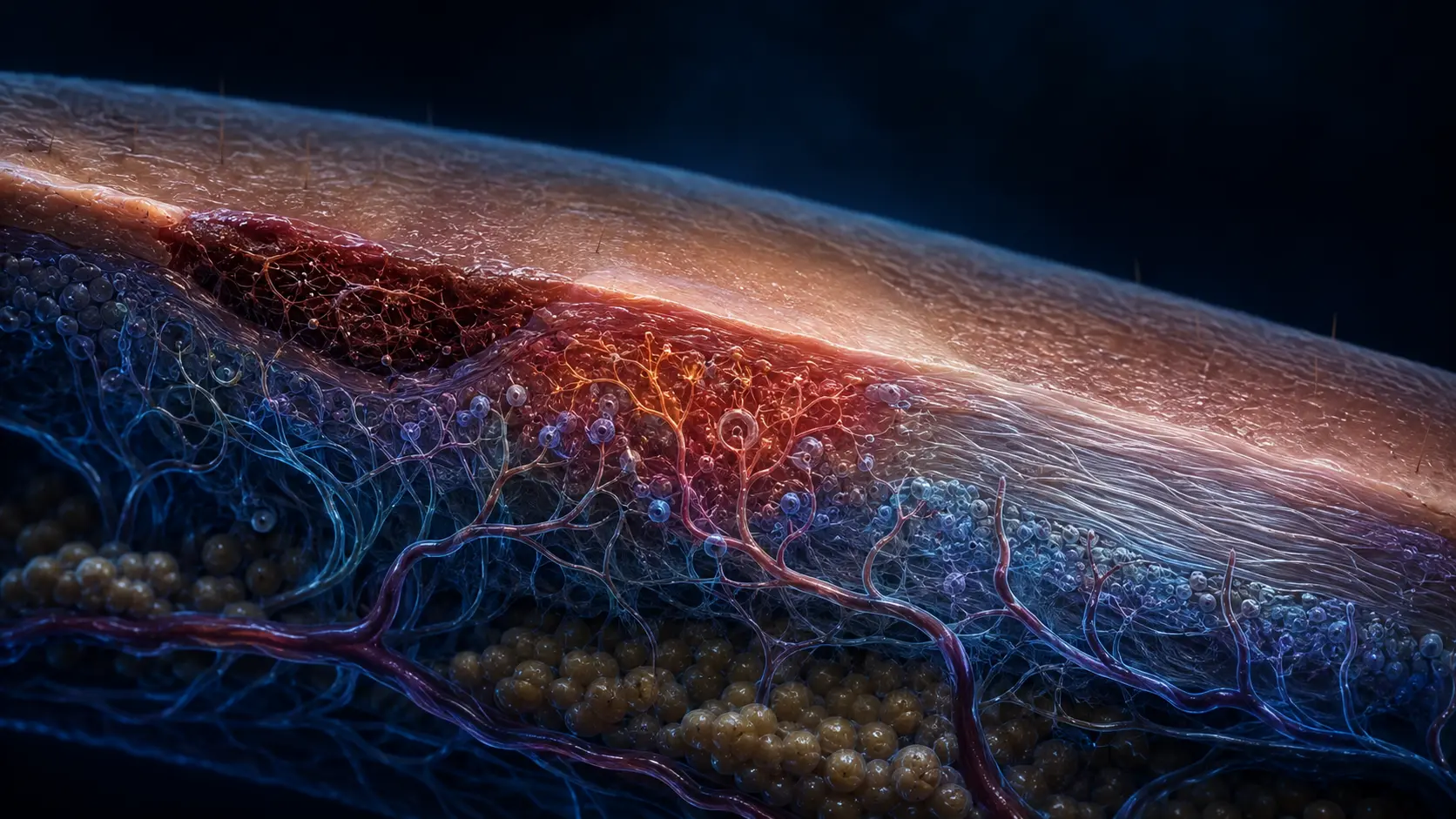
During remodeling, early matrix is degraded and replaced. Collagen fibers thicken, align with tension and form additional cross-links. Vessels regress, so a maturing scar often becomes paler and flatter.
The repair gains strength but usually never reproduces uninjured skin exactly. Collagen bundles are more parallel, elastic architecture differs, and appendages such as follicles and sweat glands may be absent in the scar core.
Mechanical tension, wound depth, genetics and sustained inflammation influence whether a scar stays narrow or becomes hypertrophic or keloid. Those raised scars have distinct biology and should not be reduced to one universal healing error.
How repair trades regeneration for speed
A clot stops leakage and provides a provisional matrix. Immune cells clean the site while surface cells, vessels and fibroblasts rebuild a continuous barrier.
Collagen-rich tissue is then remodeled under mechanical load. The patch becomes stronger and less cellular, but it preserves a repair pattern rather than the original microscopic blueprint.
A current mechanistic review follows immune cells and local signals across skin wound healing.
Why it matters
The overlapping model explains why a wound can look closed while deeper tissue is still remodeling.
It also shows why scar formation is usually a successful emergency repair, even though it falls short of complete regeneration.
A scar is repaired skin, not restored skin.
The body closes the barrier quickly with collagen-rich tissue, then remodels it for months without fully recreating the original architecture.
Research behind this story
We link to the primary study or an authoritative indexed review wherever possible. Caveats in the text reflect the limits of that evidence.
01