Quick answer
Blood clots through hemostasis. A damaged vessel narrows, and exposed structures beneath its lining capture platelets with help from von Willebrand factor. The platelets activate, change shape and recruit more platelets to form a temporary plug. At the same time, clotting reactions generate thrombin, which converts soluble fibrinogen into strands of fibrin. The fibrin mesh stabilizes the platelet plug while tissue repair begins. Anticoagulant pathways limit the reaction, and fibrinolysis later removes the clot. A clot forming inside an uninjured vessel is thrombosis, not normal wound sealing.
Blood must obey two opposite rules. It has to remain fluid through thousands of kilometers of vessels, yet it must become solid enough to seal a damaged vessel within minutes. If either rule dominates, the result can be dangerous bleeding or an unwanted blockage.
The solution is hemostasis, a tightly localized sequence involving the vessel wall, platelets, soluble clotting proteins and systems that restrain and later dismantle the repair. A clot is not simply blood drying. It is an engineered response assembled where the vessel lining has been disrupted.
The vessel wall changes the rules at the injury site
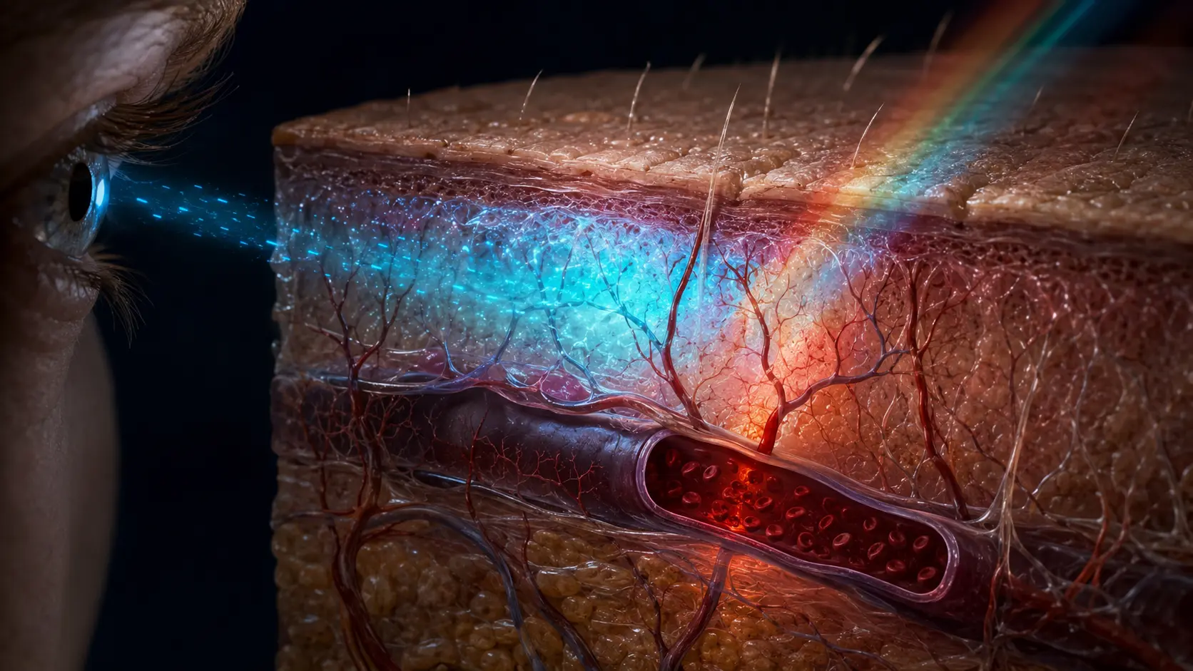
An intact endothelium normally presents a smooth, actively anti-clotting surface. It separates flowing blood from collagen and tissue factor in deeper layers and releases signals that discourage unnecessary platelet activation. Injury removes that separation in one small location.
The vessel constricts to reduce local blood flow. Exposed collagen and bound von Willebrand factor give circulating platelets something they can grip, especially where shear forces are high. The response begins at the broken wall rather than everywhere blood is present.
That localization matters. Clotting proteins circulate largely as inactive precursors, platelets remain quiet until they encounter appropriate signals, and healthy endothelium suppresses spread. Hemostasis works because activation is concentrated while multiple brakes operate around it.
Platelets build the fast temporary plug
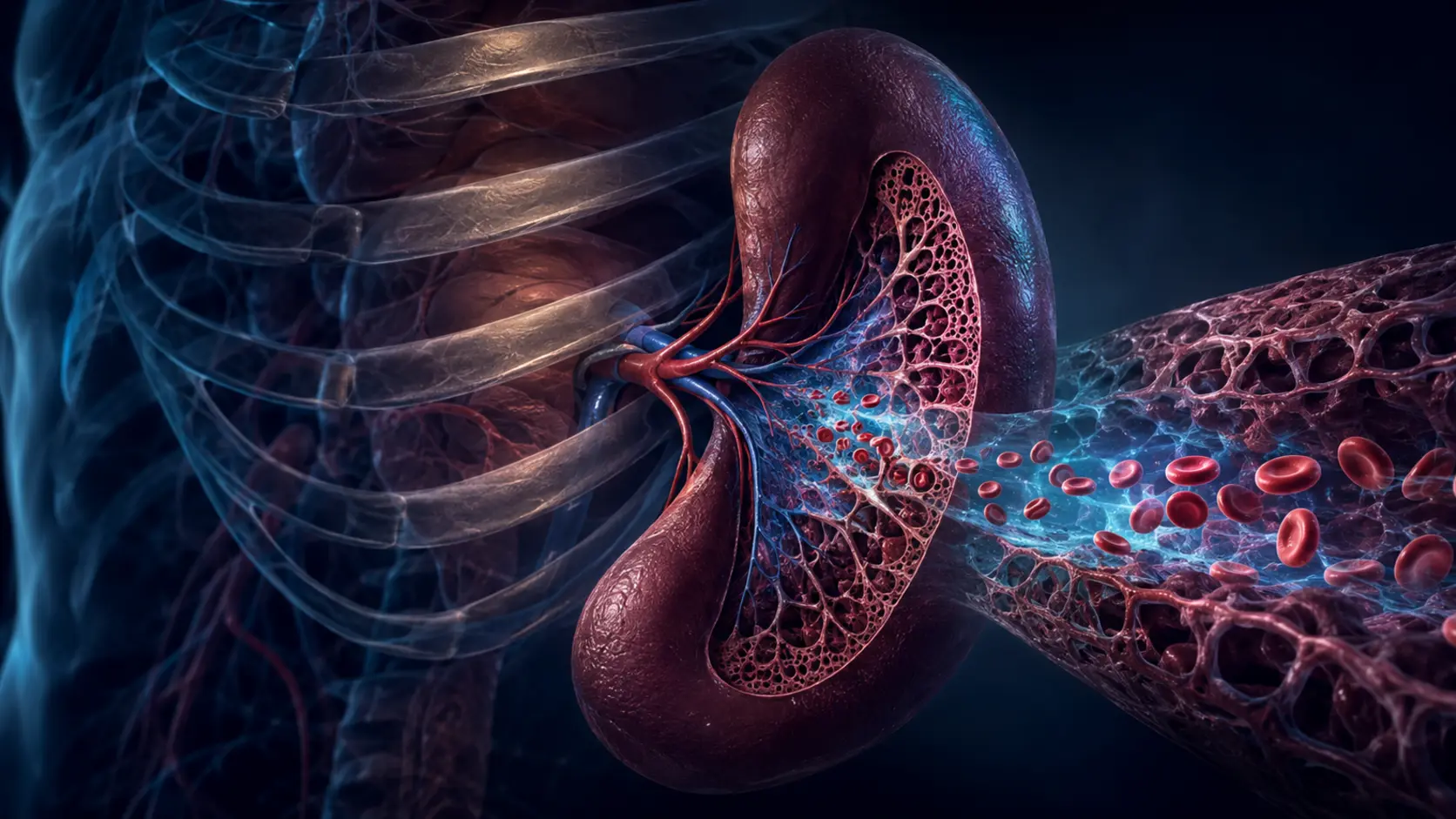
Platelets are small cell fragments produced from megakaryocytes in bone marrow. When they adhere at an injury, they flatten, extend projections and expose receptors and membrane surfaces that support further activation. They also release molecules that recruit and activate nearby platelets.
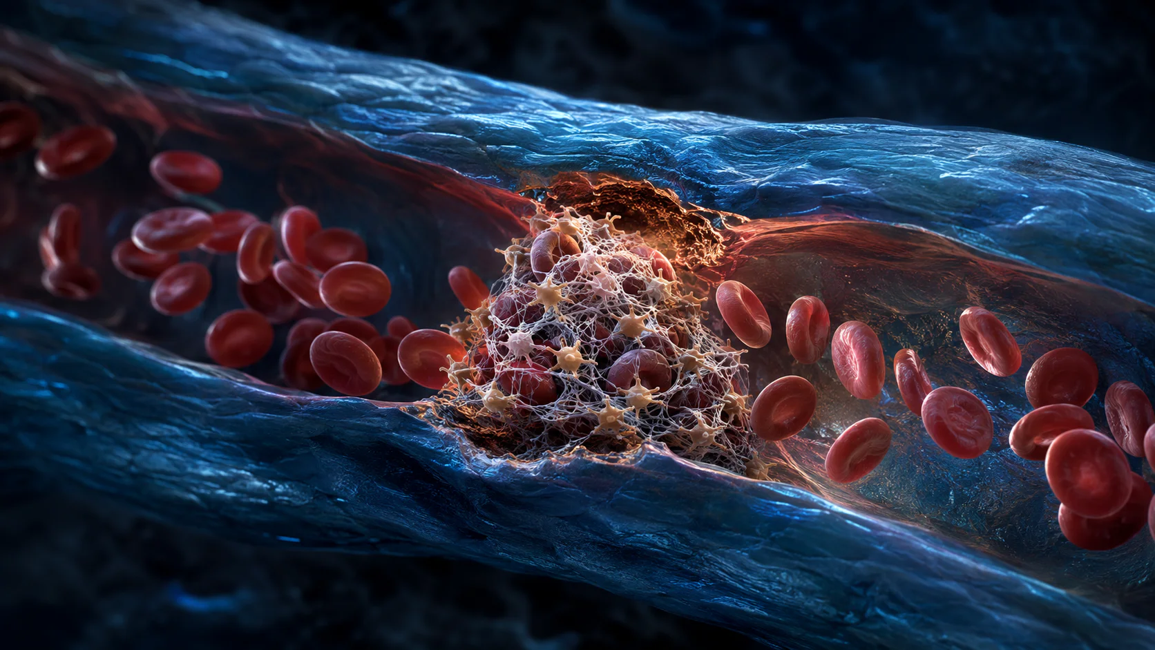
Fibrinogen can bridge activated platelets through receptors on their surfaces, allowing aggregation. This primary platelet plug can slow or stop bleeding from a small injury quickly, but it is not yet the strongest possible seal.
Platelets do more than stack. Their membranes provide a platform for coagulation reactions, and their contraction later helps compact the clot and draw damaged edges closer. They are therefore both building blocks and organizers of the next phase.
Thrombin turns soluble fibrinogen into a reinforcing mesh
Clotting factors activate one another on cell and platelet surfaces, producing an expanding burst of the enzyme thrombin near the injury. Textbook diagrams often separate intrinsic and extrinsic cascades, but clotting in living tissue is a connected, surface-dependent network rather than two isolated ladders.
Thrombin cleaves fibrinogen, a soluble plasma protein, into fibrin units that assemble into fibers. Factor XIII cross-links those fibers, creating a mesh that stabilizes the platelet plug and traps cells. The result is the firm red clot familiar at a wound.
Thrombin also amplifies platelet and factor activation, so the response accelerates once it is established. Natural anticoagulant systems—including antithrombin, the protein C pathway and tissue factor pathway inhibitor—help keep that amplification from escaping the damaged region.
A successful clot is temporary
As the vessel heals, the clot contracts and is remodeled. Plasmin, generated from plasminogen, cuts fibrin into soluble fragments in a process called fibrinolysis. Removal is controlled so the seal is not lost before repair is ready.
The same machinery can become harmful when activated in the wrong place. A thrombus inside a vein or artery can obstruct flow or break apart and travel. That is biologically different from a protective clot sealing an external cut, even though platelets and fibrin participate in both.
Unexplained swelling or pain in one limb, sudden chest pain, difficulty breathing, coughing blood, sudden weakness or speech trouble can be emergency warning signs. This explanation cannot assess symptoms or advise changes to aspirin, anticoagulants or other medicines.
From vessel damage to a stable seal
The damaged wall exposes adhesive material, platelets attach and aggregate, and clotting reactions generate thrombin on nearby surfaces.
Thrombin produces fibrin, which reinforces the platelet plug. Repair systems then rebuild the vessel while fibrinolysis gradually removes the temporary scaffold.
The NHLBI overview follows platelet-plug formation and fibrin reinforcement.
A physiology reference divides hemostasis into vessel constriction, platelets, coagulation and clot control.
Try it yourself
Separate a surface scab from the clot beneath it.
- Notice that a small cut usually stops bleeding before a dry scab is fully visible.
- The early seal is platelets and fibrin; the later scab also contains dried material at the surface.
- Leave the site undisturbed and follow normal wound-care guidance rather than testing the seal.
Bleeding that is severe, will not stop with appropriate pressure, or occurs while taking blood-thinning medicine can need urgent medical care.
Why it matters
Hemostasis shows how biology solves a control problem: create a strong positive-feedback response at one point while preventing it from spreading through the circulation.
The distinction between protective clotting and thrombosis also explains why medicines that reduce harmful clots can increase bleeding risk. The same system is being shifted, not switched off in only one location.
A blood clot is a local platelet plug reinforced with fibrin.
Vessel injury starts the assembly, natural brakes contain it, and fibrinolysis removes the scaffold after repair advances.
Research behind this story
We link to the primary study or an authoritative indexed review wherever possible. Caveats in the text reflect the limits of that evidence.
01