Quick answer
Most broken bones heal by building a temporary bridge across the fracture. A blood clot and inflammatory response first clear damaged tissue and release repair signals. Cells from the periosteum, marrow and surrounding tissues then form a soft callus rich in fibrous tissue and cartilage. Osteoblasts replace that callus with mineralized woven bone, creating a hard callus. Over months and sometimes years, osteoclasts and osteoblasts remodel the bulky bridge into stronger, better-organized lamellar bone. Alignment, blood supply, stability, fracture location, age, smoking, nutrition, infection and disease can all change how well and how quickly this happens.
A broken bone begins healing before a cast is fully set. Torn blood vessels create a clot around the fracture, immune cells enter, and signals from the damaged marrow, bone lining and surrounding soft tissue recruit cells that can rebuild the gap.
The familiar four-stage description—inflammation, soft callus, hard callus and remodeling—is useful, but the stages overlap. Repair also changes with the amount of movement at the break. The body can bridge a moderately stable gap with cartilage and woven bone, while rigidly stabilized surfaces may heal more directly.
A fracture begins as both a structural injury and a wound
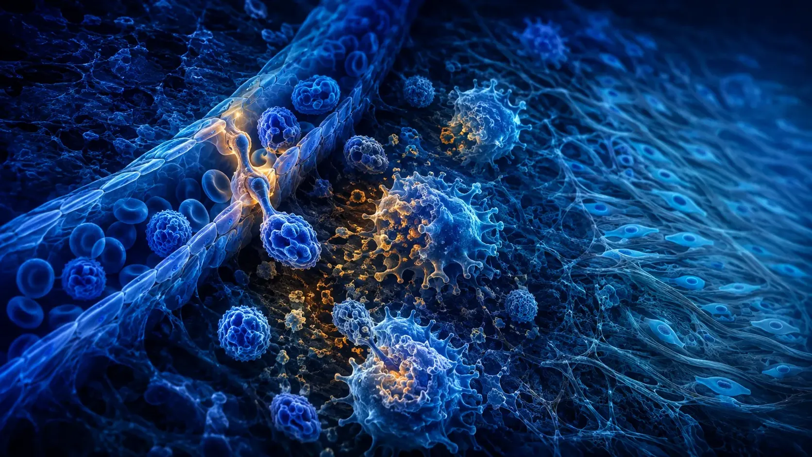
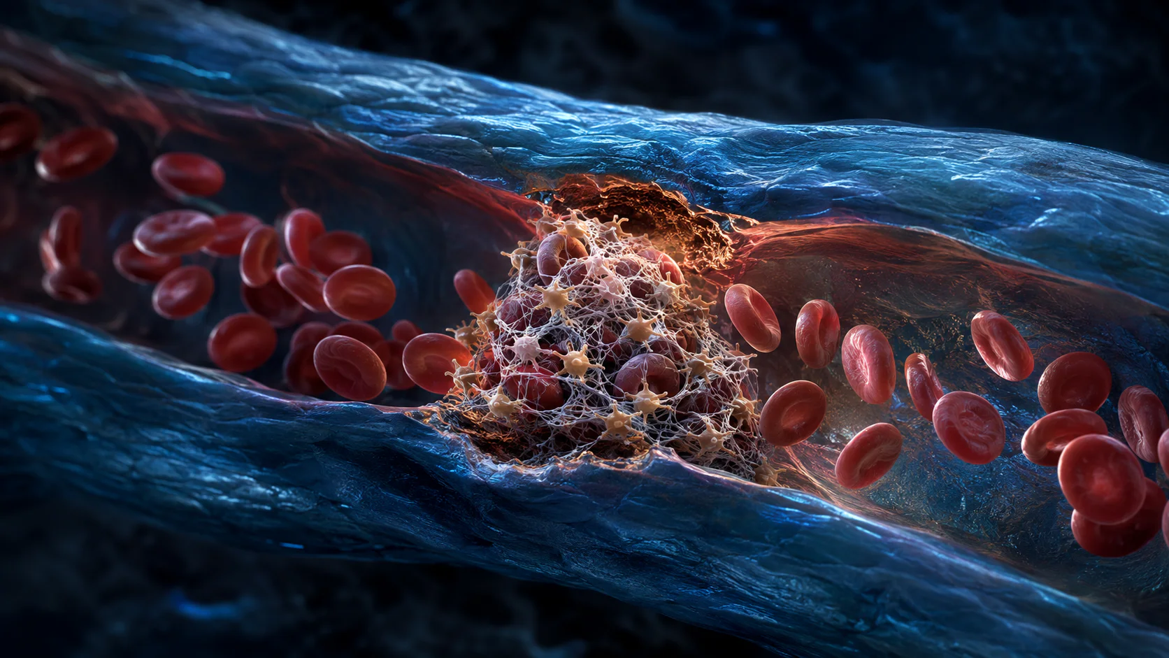
Bone contains blood vessels and living cells, so a fracture tears more than mineral. Blood collects around the break and coagulates into a hematoma. Low oxygen, damaged matrix and dying cells release molecular alarms that activate platelets, complement and immune cells.
Neutrophils arrive early, followed by macrophages and other immune populations. They remove debris and produce signals that recruit blood-vessel cells and mesenchymal stromal cells. Inflammation is therefore not waste heat around repair; a brief, controlled inflammatory phase helps organize what comes next.
Too little or too much can both be problematic. Severe soft-tissue damage, impaired circulation, infection or persistent systemic inflammation can disrupt the transition toward rebuilding. The phases are not switches that turn on one at a time—cleanup, vessel growth and tissue formation overlap from the start.
A soft callus stabilizes a gap that cannot yet carry normal load
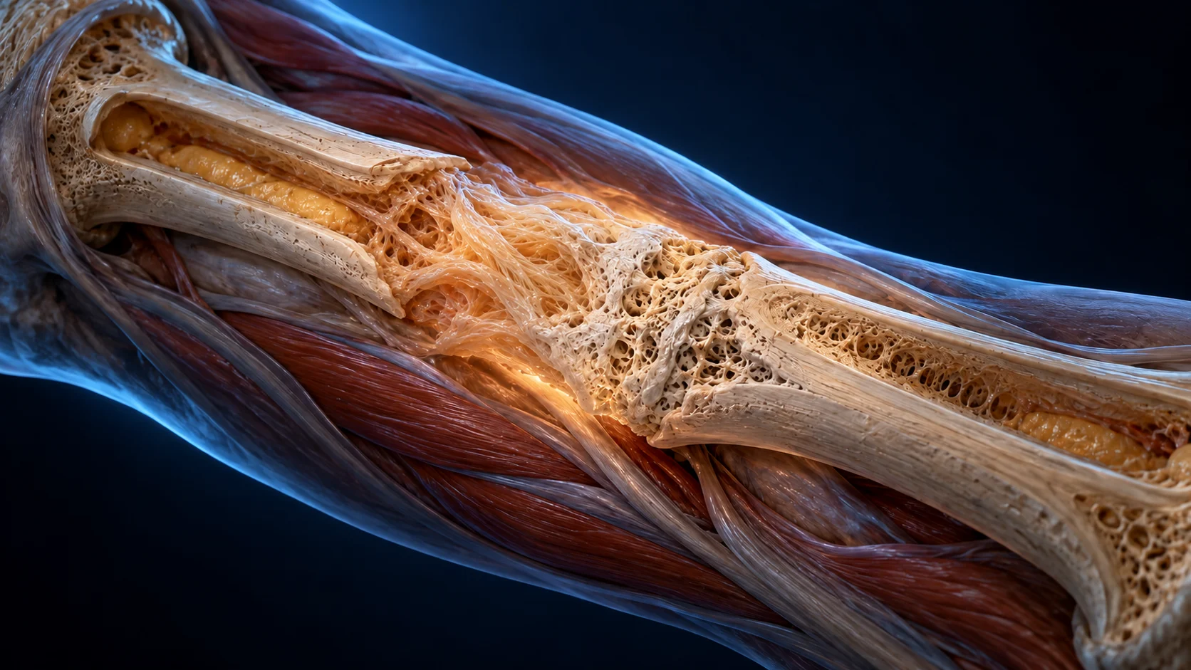
Cells from the periosteum, endosteum, marrow and nearby tissue proliferate around the fracture. Where strain and oxygen conditions favor it, some become chondrocytes and produce cartilage. Elsewhere, osteoblast-lineage cells can begin forming bone directly. The combined fibrous and cartilaginous bridge is called the soft callus.
Cartilage is useful because it can tolerate an environment that is not yet ready for mature bone. As new vessels grow into the callus, cartilage cells enlarge and the matrix changes. This resembles parts of endochondral ossification, the process that also builds many bones during development.
The callus is often larger than the original bone shaft. That apparent excess spreads force across a vulnerable region. It is biological splinting, not the final shape. Immobilization and fixation aim to keep strain within a range that allows the bridge to organize instead of repeatedly tearing.
Woven bone replaces the temporary bridge
Osteoblasts deposit collagen-rich osteoid, which mineralizes into woven bone. Woven bone is produced relatively quickly and has a disorganized fiber pattern compared with mature lamellar bone. Speed matters at this stage: the fracture needs a continuous mineralized bridge before it can be refined.
Osteoclasts and related cells help remove calcified cartilage and reshape surfaces while new bone is laid down. Blood vessels are essential because active cells need oxygen, nutrients and routes for signaling. This is why damage to the local circulation can delay union even when the bone ends appear close on an X-ray.
Clinical union does not require every trace of the fracture line to vanish. Doctors combine imaging with pain, stability, location and the demands placed on the limb. A radiograph is a two-dimensional snapshot of an evolving three-dimensional structure, so healing decisions cannot be reduced to one calendar date.
Remodeling trades fast bone for well-organized bone
Once the fracture is bridged, mechanical loading helps guide a slower remodeling phase. Osteoclasts remove selected regions of woven bone, and osteoblasts replace them with lamellar bone whose collagen layers are better organized for repeated stress.
The marrow cavity and outer contour can gradually become less bulky. Wolff's law is often summarized as bone adapting to load, but adaptation is not an instruction to stress an unhealed fracture. The timing and amount of weight bearing depend on the fracture and its treatment.
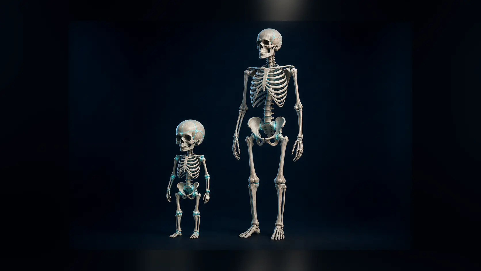
Remodeling can continue long after pain improves and a person returns to ordinary activity. Children often reshape some deformities more effectively than adults because growth remains, but that capacity varies by bone, age and the direction of the deformity. Good alignment still matters.
Not every fracture makes a large callus
The callus pathway is called secondary or indirect bone healing. It is common when a fracture has relative stability: movement is limited but tiny strains remain. The tissue passes through fibrous and cartilaginous intermediates before a hard bridge forms.
With near-anatomical alignment and very rigid fixation, some fractures can undergo primary or direct healing. Cutting cones led by osteoclasts cross the fracture line, and osteoblasts refill the channels with organized bone. A large external callus may be absent.
Neither pathway is universally superior. Surgeons choose casts, nails, plates, screws or other strategies according to the fracture pattern, soft-tissue condition and person. The mechanical environment has to match the intended biological route while preserving circulation and function.
Healing slows when biology or mechanics lose the balance
A fracture may heal slowly if the gap is too unstable, the bone ends are widely separated, blood supply is poor or infection is present. Smoking is a well-established risk factor for delayed union and nonunion. Diabetes, some medications, malnutrition, advanced age and severe injury can also influence repair, but their effects differ among people and fracture types.
A delayed union is taking longer than expected; a nonunion has stopped progressing toward healing and may need further treatment. Those are clinical judgments, not labels that can be assigned from persistent soreness alone. New deformity, increasing pain, fever, drainage, numbness, a pale or cold limb, or a cast that feels dangerously tight warrants prompt medical advice.
This article explains the biology, not an individual recovery schedule. Follow the weight-bearing, wound-care and follow-up instructions from the treating team. Feeling better does not prove that the new bridge is ready for an unrestricted load.
From broken ends to load-bearing bone
A hematoma and short inflammatory response clear damage and recruit repair cells. Fibrous tissue and cartilage form a stabilizing soft callus, which is progressively vascularized and replaced by woven bone.
The hard callus restores continuity quickly. Slower remodeling then replaces disorganized bone, reshapes the bridge and aligns tissue with the forces the bone normally carries.
A major review explains how immune activity, blood vessels, fixation and cell lineages shape fracture repair.
Recent work maps the overlapping immune and stromal cell phases of callus formation.
Why it matters
Fracture healing shows that inflammation, mechanics and regeneration cannot be separated. Cells respond not only to chemical signals but also to movement, strain and blood flow.
The callus is intentionally temporary. Successful repair builds a fast bridge first and improves it later, which explains why symptoms, X-rays and mechanical strength recover on different schedules.
A broken bone heals by bridging, replacing and remodeling.
Inflammation recruits repair cells, a soft callus stabilizes the gap, woven bone creates continuity and months of remodeling refine the result.
Research behind this story
We link to the primary study or an authoritative indexed review wherever possible. Caveats in the text reflect the limits of that evidence.
01