Quick answer
Bruises change color because blood has escaped damaged vessels and its components are gradually dismantled in tissue. Early red and purple tones come mainly from hemoglobin-rich blood and changes in its oxygen state, filtered through skin. Macrophages clear red cells; heme oxygenase converts heme into green biliverdin, and biliverdin reductase produces yellow bilirubin. Iron can be stored in ferritin or hemosiderin as cleanup continues. Depth, skin tone, blood flow, lighting and overlapping pigments alter what is visible, so color alone cannot reliably determine the exact age of a bruise.
A bruise can look red on the first day, purple or blue soon after, then acquire green and yellow edges before fading. The sequence resembles a clock, but it is not accurate enough to date an injury from color alone.
Under intact skin, small vessels have leaked blood into tissue. The changing palette records chemistry, cleanup and light passing through layers of skin—not one pigment simply losing saturation.
A bruise begins outside the circulation
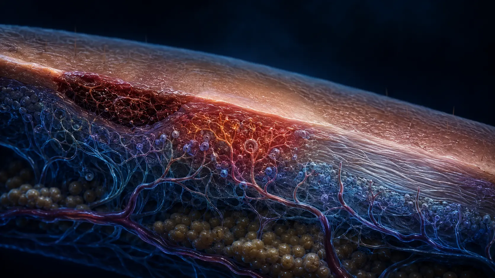
Blunt force can rupture capillaries and small veins without breaking the skin. Blood enters the extracellular space, where red cells and plasma encounter collagen, fat and resident immune cells rather than the smooth interior of a vessel.
Platelets and clotting proteins help limit continued leakage. Swelling and tenderness come from tissue injury, pressure and inflammatory signaling. The visible area may expand or shift because blood and fluid move through tissue planes.
The color seen at the surface depends on depth. A deep collection can remain faint or appear later, while a shallow one may look intense soon after injury.
Hemoglobin dominates the early palette
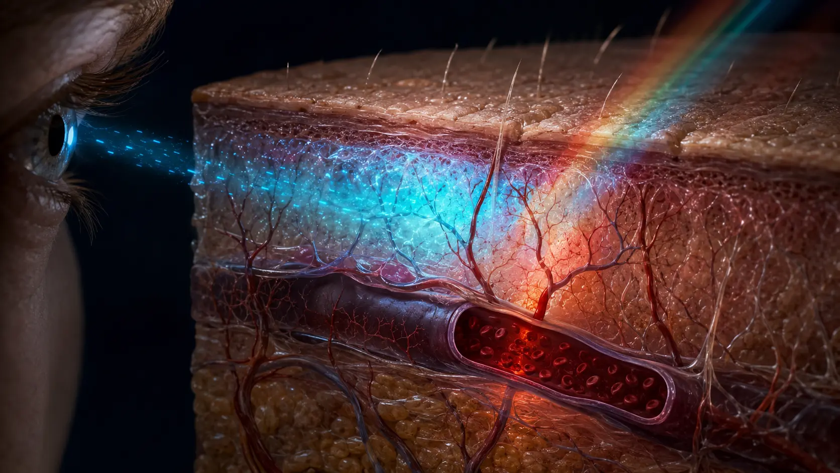
Freshly leaked blood contains red cells packed with hemoglobin. Oxygenated and deoxygenated forms absorb light differently, while the skin above scatters and filters that light. The result can range from red to dark purple, blue or nearly black.
Bruises do not literally become blue because the blood turns blue; human blood remains red. The blue appearance is an optical effect created by absorption, depth and surrounding tissue, much like the reason superficial veins can look blue.
As red-cell membranes fail, hemoglobin is exposed to enzymes and phagocytic cells. That begins a controlled disposal pathway because free heme and iron can be chemically reactive.
Heme breakdown creates green and yellow products
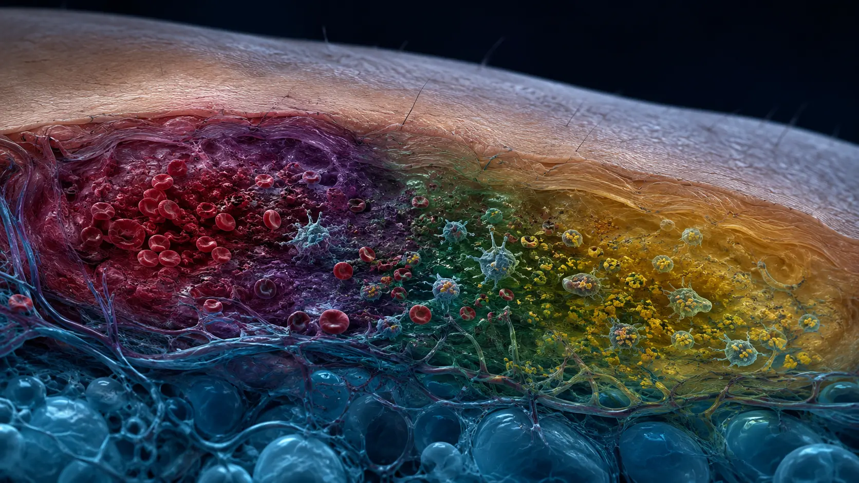
Macrophages engulf damaged red cells and hemoglobin. Heme oxygenase opens the heme ring, releasing iron and producing biliverdin, a green pigment. Biliverdin reductase then converts biliverdin into bilirubin, which is yellow.
These pigments overlap rather than appearing as four clean stages. Green or yellow may first become obvious at an edge where concentrations, depth and clearance differ from the center. Hemosiderin, an iron-storage complex, can contribute brown tones in some resolving or repeated bleeding.
The body transports and processes breakdown products while lymphatic drainage removes fluid and debris. The bruise fades when the concentration of light-absorbing material falls below what the eye can distinguish through the skin.
Color cannot reliably date a bruise
Ageing charts imply a universal progression by day, but controlled studies find wide variation and poor agreement among observers. A yellow bruise is generally not brand new, yet its exact timing cannot be assigned safely from a photograph or visual examination alone.
Location, injury force, depth, age, medications, vascular fragility and skin pigmentation all change appearance. Camera white balance and room lighting add further uncertainty, which matters in clinical and forensic interpretation.
Unexplained frequent bruising, very large bruises, bleeding elsewhere, severe pain or bruising after a significant injury merits medical advice. Normal color change explains healing chemistry; it does not explain why excessive bleeding occurred.
How trapped blood becomes a fading bruise
Vessel damage releases red cells into tissue. Clotting limits the leak while immune cells begin removing damaged material.
Hemoglobin is dismantled into biliverdin, bilirubin and iron-containing products. Drainage and cellular clearance progressively reduce the pigment load.
A review of bruise biology details the transition from hemoglobin to biliverdin, bilirubin and iron-storage products.
Why it matters
The color sequence connects everyday repair with the same heme-processing chemistry used throughout the body.
Its variability is equally important: visually plausible patterns are not precise forensic clocks.
A bruise fades by changing chemistry, not by simple dilution.
Blood pigments are converted and cleared in overlapping stages, producing a shifting mix of red-purple, green, yellow and sometimes brown.
Research behind this story
We link to the primary study or an authoritative indexed review wherever possible. Caveats in the text reflect the limits of that evidence.
01