Quick answer
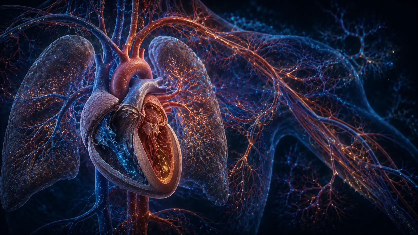
Blood circulation is a closed transport system organized into two loops. The right heart receives oxygen-poor blood from the body and pumps it through pulmonary arteries to the lungs. Gas exchange adds oxygen and removes carbon dioxide. Pulmonary veins return oxygen-rich blood to the left heart, which pumps it through the aorta into systemic arteries. Arterioles distribute flow among organs, capillaries exchange gases, nutrients, water and wastes with tissues, and veins return blood to the heart. Valves limit backflow, while pressure, vessel diameter, breathing and muscle activity help maintain movement.
Place two fingers at your wrist and the pulse feels like one repeated event. Inside the chest, that beat coordinates two pumps, four valves and a transport network whose vessels range from large arteries to capillaries narrower than a hair.
Circulation works because blood moves down pressure gradients. The heart creates those gradients, valves keep flow predominantly forward, and elastic vessels smooth each pulse into continuous delivery. The route is stable anatomy; its moment-to-moment settings change with the body's needs.
One circuit contains two different loops
Blood returning from the body enters the right atrium through the venae cavae. It crosses the tricuspid valve into the right ventricle, then passes the pulmonary valve into the pulmonary arteries. These arteries are unusual because they carry blood that has released much of its oxygen.
In the lungs, vessels divide until capillaries lie beside microscopic alveoli. Oxygen diffuses into blood while carbon dioxide moves toward alveolar air. Pulmonary veins carry the refreshed blood to the left atrium, across the mitral valve and into the left ventricle.
The left ventricle ejects through the aortic valve. Its wall is thicker because the systemic loop has far greater resistance than the short lung circuit. Both ventricles normally move about the same volume over time; a persistent mismatch would make blood accumulate in one loop.
Pressure starts flow; resistance distributes it
When ventricles contract, their pressure rises and outlet valves open. During relaxation, ventricular pressure falls, outlet valves close and inlet valves allow filling. Heart sounds mainly reflect valve closure and the vibrations that follow, not rigid doors snapping shut.
Arteries stretch during ejection and recoil between beats, helping sustain flow during diastole. Smaller arterioles contain smooth muscle that changes their diameter. They are therefore major controllers of resistance and of how much blood reaches each organ.
Flow equals pressure difference divided by resistance is a useful model, but living vessels are elastic and actively regulated. Local metabolites, nerves and hormones redirect output during exercise, digestion or temperature change without giving every organ its own pump.
Capillaries trade material with tissues
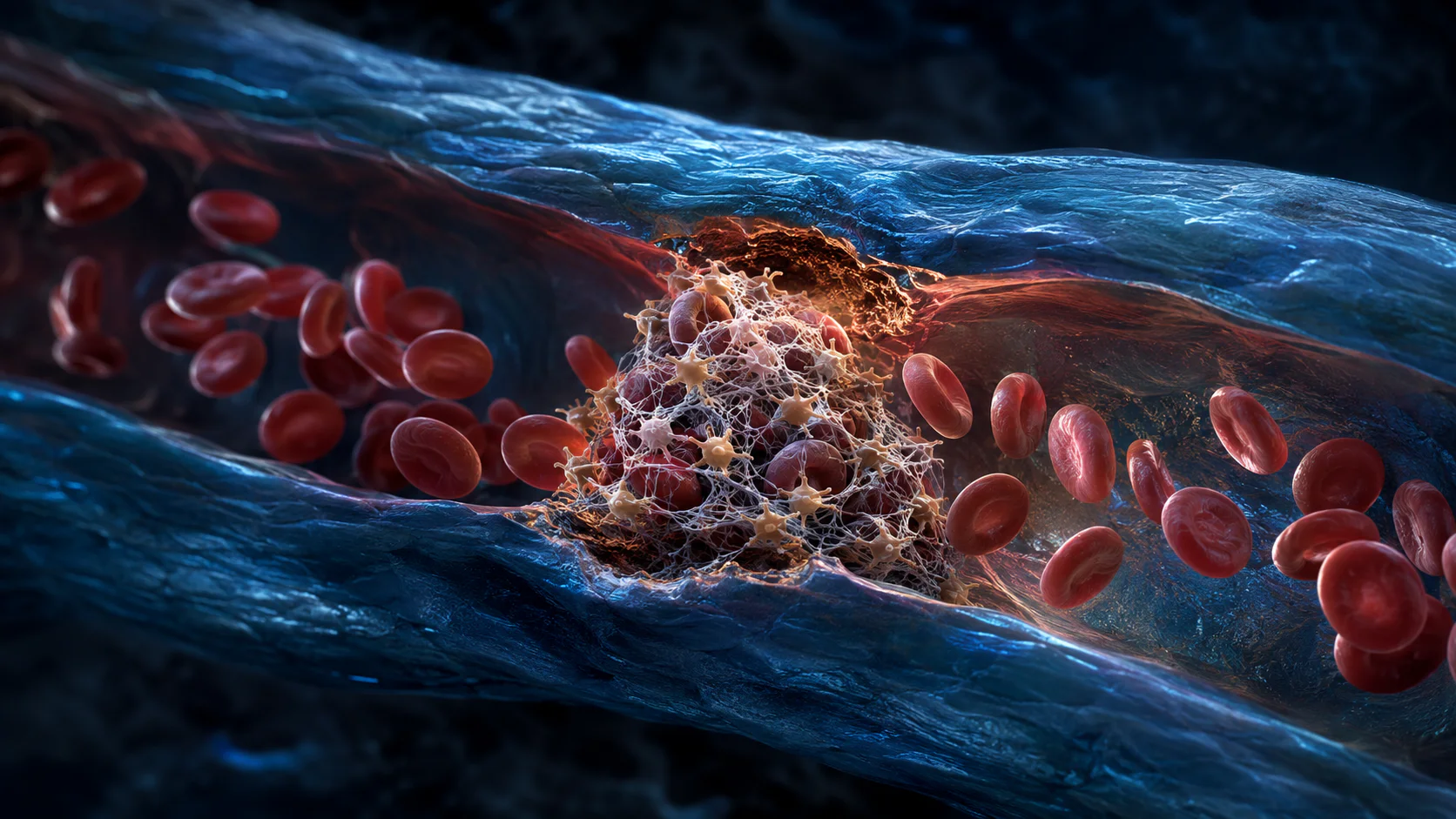
Capillary walls are usually one cell layer thick. Oxygen and carbon dioxide cross mainly by diffusion, while water and dissolved substances move according to pressure, permeability and concentration. Red cells generally remain inside intact vessels.
Not every capillary receives the same flow at every moment. Active muscle can gain more, digestive organs receive more after a meal, and skin flow changes with temperature. The broad principle is established; the precise distribution is continuously adjusted.
Some fluid leaves capillaries for spaces between cells. Most returns downstream, while lymphatic vessels collect excess and eventually return it to blood. Circulation is therefore coupled to a second fluid network rather than operating as isolated plumbing.
Return flow uses muscles, breathing and veins
Systemic veins operate at low pressure and hold much of the blood volume. Many limb veins contain valves. Contracting skeletal muscles compress them, and those valves limit backward movement, turning ordinary motion into a venous pump.
Breathing changes pressure across the chest and abdomen, assisting return toward the right atrium. Sympathetic nerves can also constrict veins during exercise or stress. These mechanisms help explain why prolonged immobility changes venous flow.
A resting and an exercising circulation use the same route but different settings. Heart rate, stroke volume and vessel tone shift together. Chest pain, fainting or sudden breathlessness cannot be interpreted from this overview and need medical evaluation.
The route of one red blood cell
A cell returning from tissue enters the right heart, crosses lung capillaries, returns to the left heart and enters the systemic arterial tree.
Pressure gradients drive each segment, valves preserve direction and vessel diameter determines how output is shared.
The American Heart Association traces blood through the chambers, valves, lungs and body.
Why it matters
The two-loop model explains why pulmonary and systemic arteries carry different oxygen levels without breaking the artery definition.
It connects the heart to lungs, kidneys, lymph vessels and skeletal muscles rather than treating circulation as one organ's job.
The heart creates gradients; vessels turn them into delivery.
Two synchronized loops refresh blood in the lungs, distribute it through the body and return it for the next beat.
Research behind this story
We link to the primary study or an authoritative indexed review wherever possible. Caveats in the text reflect the limits of that evidence.
01