Quick answer
Brain tissue itself cannot feel pain because it lacks nociceptors. Headaches can still be intense because pain-sensitive structures surround and connect to the brain. These include the meninges, parts of cranial blood vessels, the scalp, neck muscles, eyes, sinuses and cranial nerves. Much intracranial pain travels through trigeminal nerve pathways into the brainstem, thalamus and cortical networks that construct location, intensity and unpleasantness. Migraine is not simply a swollen blood vessel; it involves altered brain excitability and trigeminovascular signaling. A sudden severe or neurologically unusual headache requires urgent medical assessment.
Neurosurgeons can sometimes operate on exposed brain tissue while a carefully selected patient is awake enough to answer questions. That unsettling fact does not mean the head is numb. It reveals a boundary between the tissue that generates conscious pain and the tissues that can trigger it.
Brain parenchyma lacks the peripheral nociceptors that detect damaging mechanical, thermal or chemical events. The scalp, periosteum, muscles, eyes, sinuses, meninges, cranial arteries and nerves do contain pain-sensitive fibers. Their signals enter neural circuits that create the experience of a headache.
Pain starts with sensors but becomes an experience
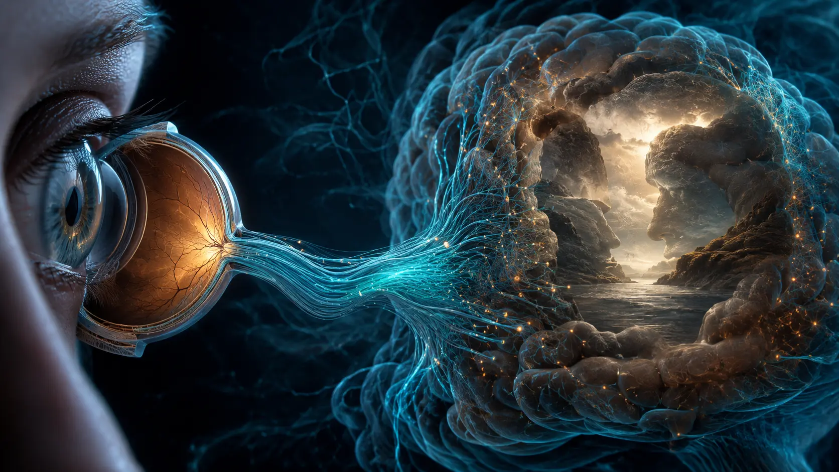
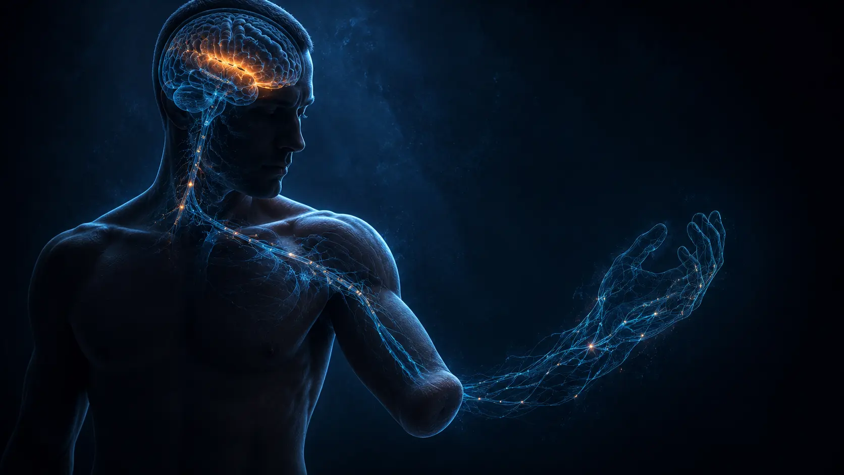
Nociceptors are sensory neurons specialized to detect potentially damaging conditions. Their endings transform pressure, temperature or injury-related chemicals into electrical signals. Those signals are nociception—the neural input associated with threat—not the complete subjective experience of pain.
The spinal cord or brainstem relays and modifies incoming activity. Thalamic and cortical networks contribute location and intensity, while insular, cingulate and other systems shape salience and unpleasantness. Attention, expectation, previous experience and state can alter the result without making the signal imaginary.
Brain tissue performs that interpretation but contains no ordinary pain endings of its own. Cutting or electrically stimulating parenchyma can affect function depending on location, yet does not produce the same local pain signal as cutting skin or dura.
The coverings and vessels around the brain are innervated
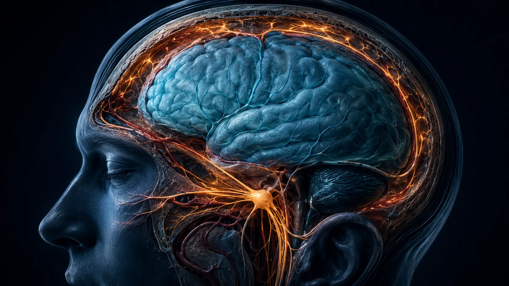
Three meningeal layers surround the brain. The outer dura and portions of cranial vasculature receive sensory fibers, many from the ophthalmic division of the trigeminal nerve. Upper cervical nerves also contribute, helping explain why head and neck pain can overlap.
Mechanical traction, inflammation and chemical mediators can activate or sensitize these afferents. Signals enter the trigeminal ganglion and brainstem trigeminocervical complex before ascending into networks that produce head pain and associated sensitivity.
The skull itself is not a simple container with one pain map. Different intracranial and extracranial tissues have different innervation, and pain can be referred. A person may feel pain at the forehead or around an eye even when the relevant activity involves deeper meningeal pathways.
Different headaches recruit different mechanisms
Migraine involves a genetically influenced nervous system, altered sensory processing and activation of the trigeminovascular system. Peptides such as CGRP participate in signaling. Blood vessels matter, but the old explanation that migraine is merely vasodilation is incomplete.
Tension-type headache is associated with pericranial tenderness and, in frequent forms, changes in central pain processing. Cluster headache has a distinct pattern involving trigeminal pain and cranial autonomic features. Infection, trauma, pressure changes and vascular disorders can generate secondary headaches through other routes.
This variety is why 'the brain has no pain receptors' does not identify the cause of one person's headache. It answers an anatomy question, not a diagnosis.
The paradox does not make severe head pain harmless
A headache that peaks suddenly within seconds or minutes, follows a significant injury, occurs with weakness, confusion, seizure, fainting, fever with neck stiffness, new visual loss or during pregnancy or postpartum can require urgent evaluation. New headaches in people with cancer or immune suppression also deserve prompt attention.
Most headaches are primary disorders rather than evidence that brain tissue is being injured. Even so, pattern, timing, accompanying symptoms, age and medical history determine risk; intensity alone cannot reliably separate causes.
The established distinction is anatomical: parenchyma lacks nociceptors while surrounding tissues are innervated. Researchers continue to refine how meningeal immune cells, vascular signaling and central sensory networks interact across migraine phases.
From a cranial tissue signal to head pain
A pain-sensitive meningeal, vascular, muscular or cutaneous ending is activated or sensitized. Trigeminal or upper cervical afferents carry the signal into the brainstem.
Ascending networks integrate the input with context, attention and state. The brain then produces the conscious location, intensity and unpleasantness of pain.
A modern review maps how meningeal afferents connect the brain's borders to migraine pain.
Why it matters
The distinction separates nociception from pain and resolves the apparent contradiction between a painless brain and painful headaches.
It also blocks a misleading conclusion: severe head pain can be medically important even though the brain's own tissue lacks pain endings.
Your brain can create pain without being able to feel its own tissue.
Head pain usually begins in innervated coverings, vessels, muscles or nerves, then becomes a conscious experience inside distributed brain networks.
Research behind this story
We link to the primary study or an authoritative indexed review wherever possible. Caveats in the text reflect the limits of that evidence.
01