Quick answer
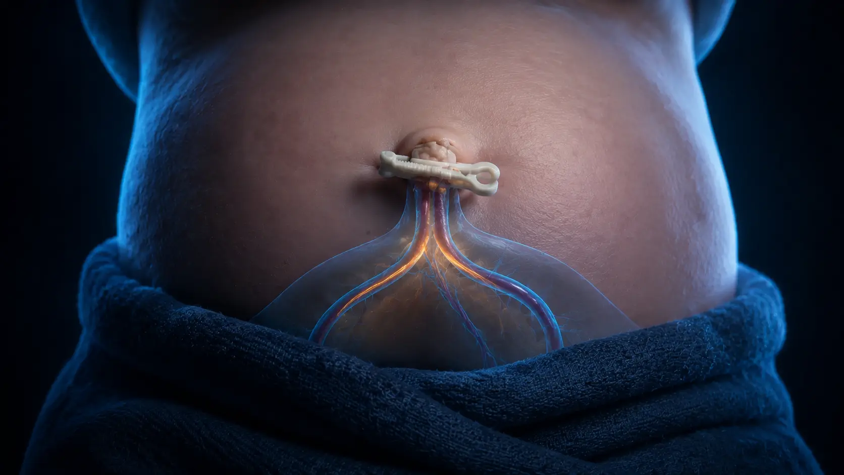
After birth, the lungs expand and pulmonary blood flow rises while the umbilical circulation is eventually clamped and separated from the placenta. Smooth muscle in the umbilical arteries constricts strongly, and flow through the umbilical vein ends. Pressure changes help close fetal shunts as circulation adopts the newborn pattern. The external cord stump dries, darkens and usually separates after one to two weeks. Internally, the umbilical vein becomes the ligamentum teres of the liver; distal umbilical arteries become medial umbilical ligaments, while proximal portions remain part of the arterial supply to the bladder.
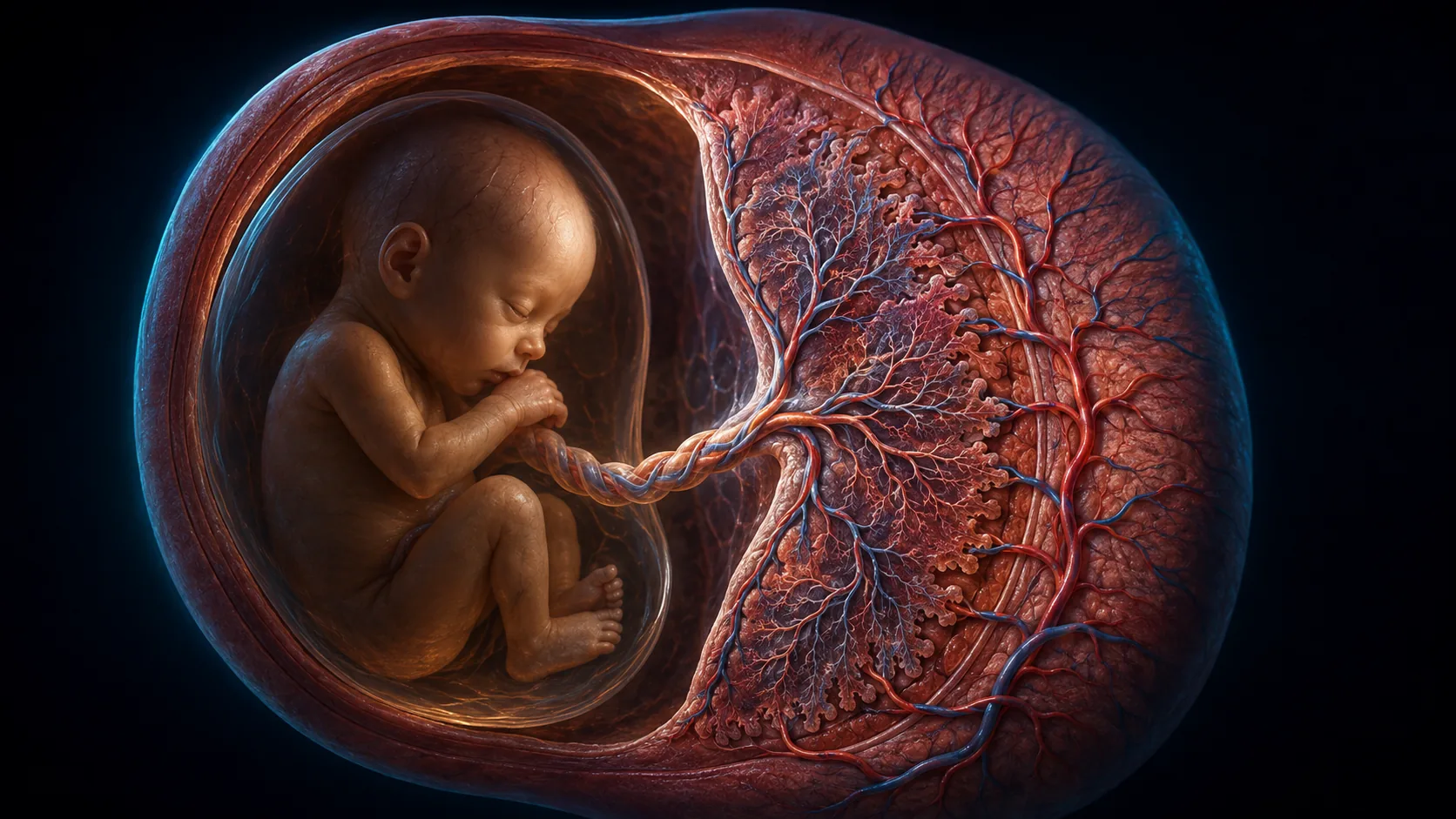
Before birth, one umbilical vein carries oxygen-rich blood from the placenta and two umbilical arteries return blood toward it. Within minutes of birth, that three-vessel circuit is no longer the newborn's route for gas exchange.
The switch is coordinated with lung aeration, changing pressures and cord clamping. Outside the body, a short stump dries and falls away; inside, the former vessels close and are remodeled into structures that remain through adulthood.
Before birth, the placenta handles gas exchange
The umbilical vein brings placental blood toward the fetus. Much of it passes through the ductus venosus toward the heart, while the two umbilical arteries branch from the fetal pelvic circulation and return blood to the placenta.
Because the lungs are fluid-filled and pulmonary resistance is high, fetal blood uses the foramen ovale and ductus arteriosus to bypass much of the lung circuit. The cord is therefore part of an entire fetal circulation, not an isolated tube.
Umbilical vessel names follow the direction of flow relative to the fetal heart. The vein carries blood toward the fetus even though its oxygen level is higher than that in the umbilical arteries.
Lung aeration and clamping change the pressure map
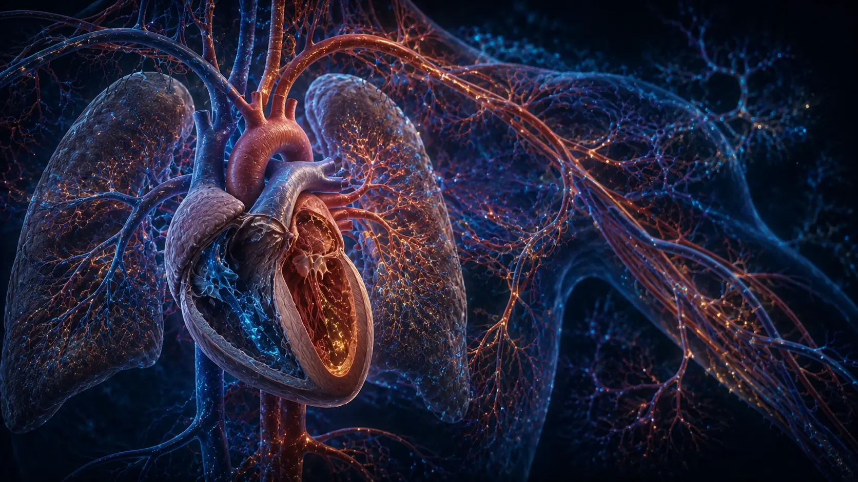
With the first effective breaths, air replaces lung fluid and pulmonary vascular resistance falls. More blood crosses the lungs and returns to the left side of the heart.
Clamping removes the low-resistance placental circulation and ends umbilical venous return. Systemic resistance rises, while increased left-atrial pressure helps press the flap of the foramen ovale toward functional closure.
The order and timing matter physiologically. This is why modern guidance considers the infant's breathing and generally supports delayed rather than routine immediate clamping when clinical circumstances allow. Individual birth decisions belong to the care team.
The vessels close first, then become anatomical remnants
Umbilical arteries contain abundant smooth muscle and constrict rapidly as temperature, oxygen and local signals change. The vein also closes, though its behavior and timing differ.
Over time, the closed umbilical vein becomes the round ligament of the liver. The ductus venosus becomes the ligamentum venosum. Distal umbilical arteries form the medial umbilical ligaments, while their proximal segments remain open as superior vesical arteries.
These adult remnants are evidence of developmental rerouting. They are not empty mistakes; they mark vessels that were essential in an earlier circulation.
The external stump separates by controlled drying
After clamping and cutting, the remaining cord loses blood flow, dries and darkens. Immune cells help create a line of separation at the skin, and the stump detaches as the umbilical area heals.
The exact day varies. Clean, dry care recommendations and local clinical protocols should be followed rather than applying substances without guidance.
Spreading redness, pus, foul odor with inflammation, fever, persistent bleeding or a newborn who seems unwell requires prompt professional assessment. A normal timeline cannot rule out infection in an individual infant.
How circulation switches at birth
Lung aeration lowers resistance in the pulmonary vessels and increases blood return from the lungs. Cord clamping removes placental flow and raises systemic resistance.
Pressure and oxygen changes promote functional closure of fetal pathways. Umbilical vessels constrict, then fibrose into adult remnants as the external stump separates.
WHO includes delayed cord clamping in essential newborn care.
Why it matters
The transition explains why breathing and cord management are physiologically connected rather than separate events.
The adult ligaments reveal a general rule of development: temporary routes can be remodeled instead of erased.
The cord becomes unnecessary because the entire circulation is rerouted.
Breathing shifts blood toward the lungs, clamping ends placental flow, and the closed vessels remodel into lasting anatomical traces.
Research behind this story
We link to the primary study or an authoritative indexed review wherever possible. Caveats in the text reflect the limits of that evidence.
01