Quick answer
Red blood cells carry oxygen mainly through hemoglobin, an iron-containing protein with four oxygen-binding sites. In lung capillaries, oxygen diffuses from alveolar air into plasma and then into red cells, where it binds hemoglobin reversibly. The oxygenated cells travel through the left heart and systemic arteries. In tissue capillaries, lower oxygen pressure draws oxygen off hemoglobin and toward cells. Higher carbon dioxide, greater acidity and warmer temperature in active tissues can reduce hemoglobin's oxygen affinity, helping unloading. The biconcave, flexible cell shape provides a large surface-area-to-volume ratio and allows red cells to squeeze through narrow capillaries. Oxygen delivery still depends on the entire system: ventilation, diffusion, hemoglobin concentration, blood flow and tissue demand all matter.
A breath brings oxygen to the lung surface, but air never travels directly to a working muscle or a thinking neuron. The final journey happens inside blood, packed into flexible cells that repeatedly load, carry and unload a gas their tissues cannot store in large amounts.
Red blood cells make that journey possible because they are filled with hemoglobin. This protein binds oxygen where its partial pressure is high, releases more of it where pressure falls and responds to the chemical conditions created by active tissue. The cell is not a sealed oxygen tank. It is a moving, adjustable delivery package.
Hemoglobin carries far more oxygen than plasma alone
A small amount of oxygen dissolves directly in plasma, but most is transported on hemoglobin inside red cells. Each hemoglobin molecule contains four heme groups. An iron atom within each heme can bind one oxygen molecule without permanently consuming it, so the same protein can load and unload oxygen over and over.
Binding is cooperative. When one oxygen molecule binds, hemoglobin changes shape in a way that makes additional binding easier. When oxygen begins to leave, the reverse structural change favors further unloading. Plotting oxygen pressure against hemoglobin saturation therefore produces a sigmoid, or S-shaped, curve rather than a straight line.
The flatter upper part helps hemoglobin remain highly saturated across the oxygen pressures normally found in the lungs. The steeper middle portion means a modest fall in tissue oxygen pressure can release a useful additional fraction. This curve is a model of equilibrium, not a fixed loading schedule for every capillary or person.
Loading begins across an extremely thin lung barrier
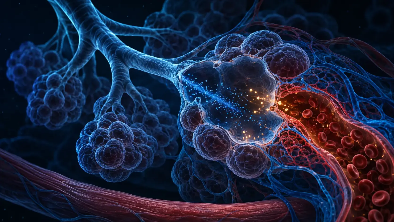
In the lungs, inhaled oxygen reaches microscopic alveoli. Oxygen partial pressure is higher in alveolar gas than in the relatively oxygen-poor blood arriving through pulmonary arteries, so oxygen diffuses across the alveolar and capillary walls, through plasma and into red blood cells.
Binding to hemoglobin keeps the freely dissolved oxygen concentration from immediately reaching equilibrium. More oxygen can continue crossing while available binding sites are filled. The newly oxygenated blood then returns through pulmonary veins to the left side of the heart and enters the systemic circulation.
A pulse oximeter estimates the percentage of hemoglobin binding sites occupied by oxygen, not the total amount of oxygen delivered to every tissue. Delivery also depends on how much hemoglobin is present and how much blood the heart moves. A saturation value cannot, by itself, describe the entire oxygen-transport system.
Tissues create conditions that favor unloading
Cells continually consume oxygen during aerobic metabolism. That lowers oxygen pressure in the fluid around them, establishing a gradient from capillary blood toward tissue. Oxygen leaves hemoglobin, diffuses out of the red cell and crosses the capillary wall along that gradient.
Active tissue also produces carbon dioxide and acids and often becomes warmer. Higher carbon dioxide and lower pH reduce hemoglobin's affinity for oxygen, a relationship called the Bohr effect. On the dissociation curve, these conditions shift the relationship toward easier unloading at a given oxygen pressure.
The effect is a tendency, not an address label that directs each oxygen molecule. Local blood flow, diffusion distance, capillary recruitment, mitochondrial demand and transit time all influence how much oxygen reaches a region. Hemoglobin chemistry works inside that larger delivery network.
The cell's shape supports rapid exchange and narrow travel
A mature human red blood cell has a biconcave disc shape: thin in the center and thicker around the rim. This geometry provides substantial membrane area relative to cell volume and keeps much of the hemoglobin-containing interior close to the surface where gases cross.
Red cells also lose their nucleus and most organelles during maturation. That leaves more internal space for hemoglobin and prevents the mature cell from using the oxygen it transports in mitochondria. It still requires energy to maintain its membrane and ion balance, relying mainly on glycolysis.
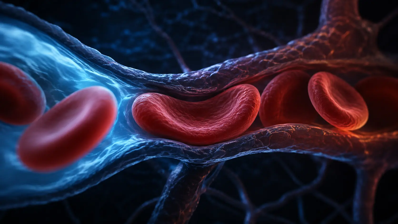
Capillaries can be narrower than an unstressed red cell. A flexible membrane and cytoskeleton let the cell bend and elongate as it passes through. Deformability supports close contact with capillary walls and continued flow; it is an essential mechanical part of delivery, not merely a visual curiosity.
Affinity can shift, but neither direction is always better
Inside red cells, 2,3-bisphosphoglycerate, usually shortened to 2,3-BPG, binds preferentially to deoxygenated hemoglobin and lowers its oxygen affinity. This favors unloading. Temperature, pH, carbon dioxide and hemoglobin type can also shift the binding relationship.
Lower affinity can help release oxygen to tissues but may make loading harder when lung oxygen pressure is very low. Higher affinity can support loading yet resist unloading. The useful balance depends on the lungs, circulation, tissues and environment; a simple rightward or leftward curve shift is not automatically beneficial.
This article describes normal transport physiology. Fatigue, breathlessness, abnormal pulse-oximeter readings or suspected anemia have many possible explanations and cannot be interpreted from the curve alone. Measurements and symptoms require appropriate clinical context.
From alveolar air to a working cell
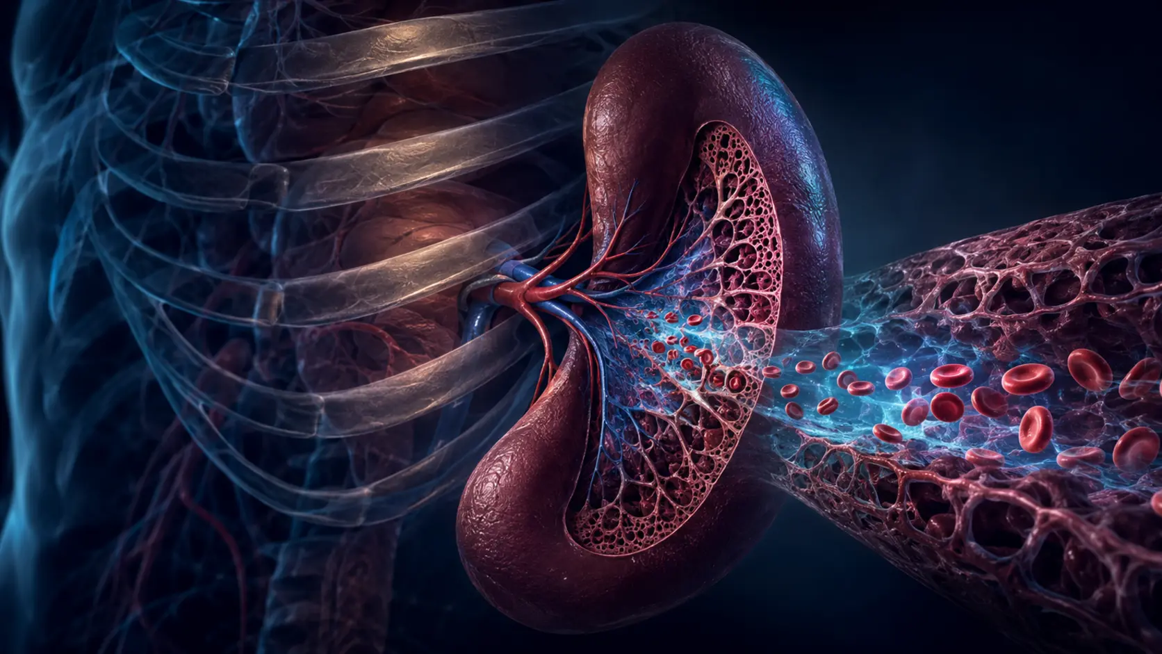
Oxygen diffuses across the lung barrier and binds cooperatively to hemoglobin inside red cells. The heart then sends those cells into systemic arteries and progressively smaller vessels.
In tissue capillaries, falling oxygen pressure and local metabolic conditions favor release. Oxygen diffuses toward cells, while the red cell bends through the microcirculation and continues toward the venous return.
NCBI's oxygen-transport overview explains why hemoglobin carries most blood oxygen while only a small fraction dissolves in plasma.
A physiology review describes how healthy cells deform under shear to pass efficiently through capillaries.
Why it matters
The mechanism separates oxygen saturation from oxygen delivery. Saturation describes hemoglobin occupancy; delivery also requires enough hemoglobin and adequate blood flow.
It also shows why red cells are more than passive bags. Protein cooperativity, small-molecule regulation, cell geometry and deformability work together across the lung and tissue sides of one cycle.
Red blood cells carry oxygen by reversible binding, not storage alone.
Hemoglobin loads oxygen where pressure is high and releases more where pressure falls and tissue chemistry lowers affinity.
Research behind this story
We link to the primary study or an authoritative indexed review wherever possible. Caveats in the text reflect the limits of that evidence.
01