Quick answer
Your pupils dilate in the dark because less retinal light shifts the autonomic balance controlling the iris. Parasympathetic activity that keeps the circular sphincter muscle contracted falls quickly, allowing that muscle to relax; sympathetic activity also activates the radial dilator muscle. The wider opening lets more light reach the retina. It helps immediately, but it is only one part of night vision: rods, cones and retinal circuits continue adapting over minutes. Pupil size also changes with viewing distance, medicines, arousal, age and individual anatomy, so a large pupil is not a simple readout of one emotion or thought.
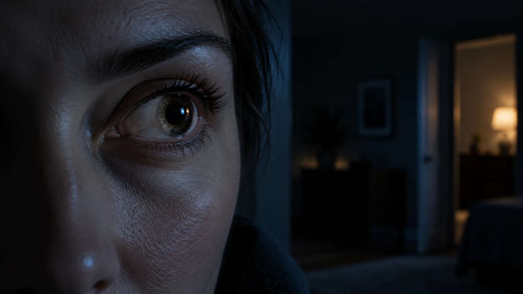
Step from a bright hallway into a dark room and the black opening at the center of each eye grows. The change is automatic: you do not decide to enlarge it, and the pupil itself is not a piece of tissue that stretches.
The moving structure is the iris. Two smooth-muscle systems change the diameter of its central opening while retinal and brain circuits measure light and adjust the command.
The pupil is an opening controlled by the iris
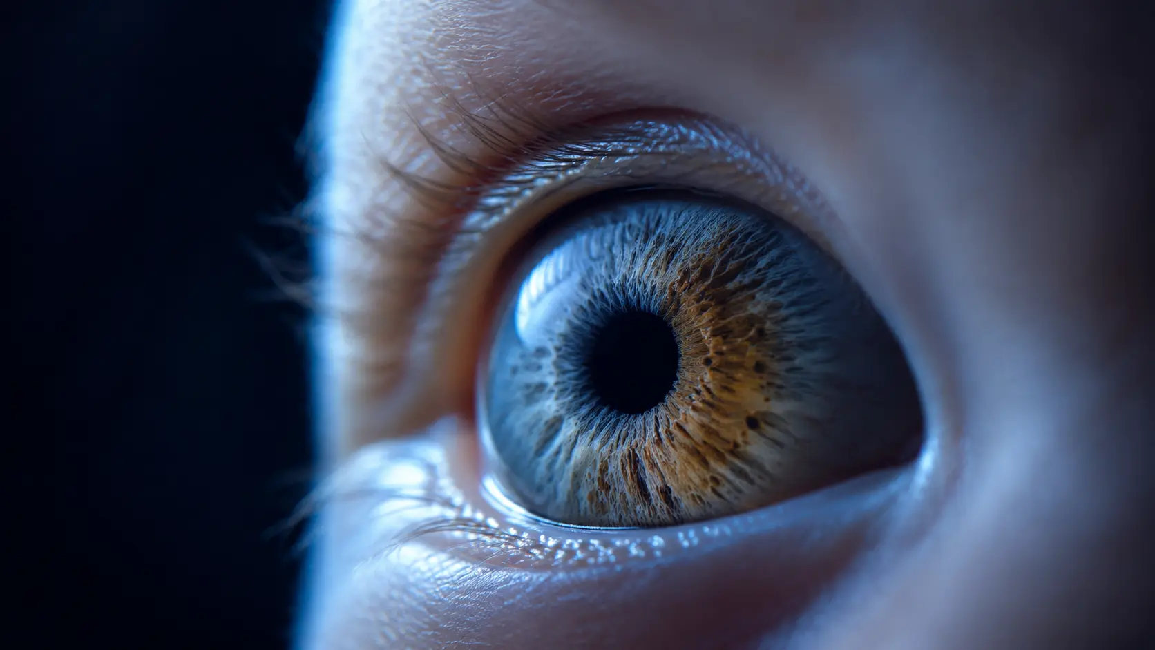
The pupil looks black because it is an aperture leading into the eye, where much of the entering light is absorbed rather than reflected back. The colored iris forms the adjustable ring around that opening. Its texture and melanin shape visible eye color, but its muscles set pupil diameter.
Circular fibers called the sphincter pupillae narrow the opening when they contract. Radially arranged fibers of the dilator pupillae pull the iris outward when they contract. Both are smooth muscles governed automatically rather than skeletal muscles under conscious control.
That opposing arrangement lets the eye respond over a wide range of illumination. Bright light favors a smaller aperture that limits retinal exposure and reduces some optical blur. Dim light favors a larger aperture that captures more photons, with a tradeoff: aberrations and depth of field can worsen as the opening grows.
The retina starts a two-sided light reflex
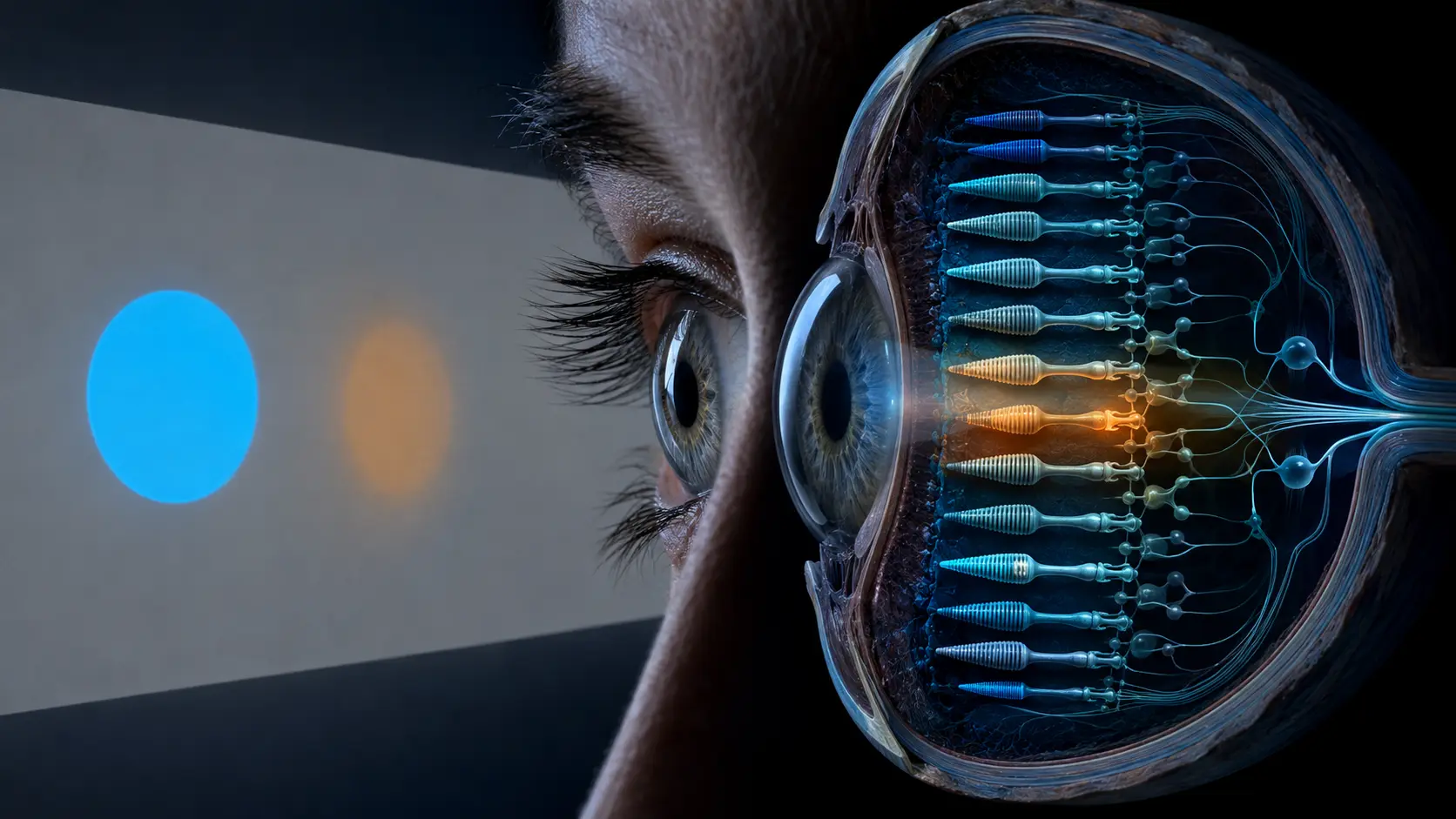
Rods and cones contribute information about illumination, and melanopsin-containing intrinsically photosensitive retinal ganglion cells help sustain the pupil's response to light. These ganglion cells also receive rod-and-cone input, so the reflex is a network response rather than the work of one photoreceptor type.
Signals travel from each retina through the optic nerve to nuclei in the midbrain. Connections then reach both Edinger–Westphal nuclei, whose parasympathetic fibers run with the third cranial nerve to the ciliary ganglion and finally the iris sphincter.
Because the midbrain distributes light information to both sides, shining light into one healthy eye normally constricts both the illuminated pupil and its partner. Clinicians call these the direct and consensual light responses. Their pattern can help localize a pathway problem, but self-testing cannot replace an eye or neurological examination.
Darkness releases a brake before the dilator adds pull
When illumination falls, retinal drive to the constricting pathway decreases. Parasympathetic output drops and the sphincter relaxes. Research on the early dilation phase indicates that this release of sphincter tone makes a major contribution to the first rapid widening.
The sympathetic pathway also matters. It descends through the brainstem and spinal cord, passes through the superior cervical ganglion and reaches the radial dilator muscle. Its contraction pulls the iris outward and helps maintain a larger pupil, particularly during darkness and arousal.
Calling dilation simply a sympathetic fight-or-flight response misses half the mechanism. In ordinary darkness, reduced parasympathetic constriction and sympathetic dilator activity work together, with their relative contributions changing over time and with context.
A wider pupil is not the same as dark adaptation
Opening the aperture increases the light available immediately, but it cannot fully explain why a dark room becomes easier to see after several minutes. That slower improvement happens largely in the retina as photopigments recover and neural circuits shift toward the rod system's high sensitivity.
Cones support color and fine detail in brighter conditions. Rods are more sensitive at low light but do not encode color the same way and provide lower spatial detail. During dark adaptation, sensitivity can keep improving long after pupil diameter has approached a new level.
This distinction explains why walking indoors from sunlight can feel nearly black even though the pupils have already begun opening. The aperture is changing, while retinal chemistry and signaling still need time to recalibrate.
Pupil size carries more than a light signal
Near focus usually constricts the pupils as part of the near response, alongside lens accommodation and inward eye movement. Arousal, surprise, effort and attention can produce smaller changes through autonomic and brainstem systems. Those influences sit on top of the strong response to ambient light.
That is why a photograph of a large pupil cannot reveal a specific emotion, attraction or lie. Lighting, viewing distance, camera flash, age, eye color, medicines and recreational drugs can all change the measurement. Scientists control those variables carefully when using pupillometry as an indirect marker of brain state.
Maximum pupil size tends to decrease with age, especially in dim conditions, and healthy people vary substantially. The two pupils may also differ slightly in some people without disease. What matters clinically is the pattern, timing and accompanying symptoms rather than a universal millimeter cutoff read from a mirror.
A sudden new difference needs context
A small, stable difference between pupils—called physiological anisocoria—can be normal. A new unequal appearance, a difference that persists, or a pupil that does not react normally deserves professional assessment because eye, nerve, medicine and brain causes can look similar without examination.
Seek urgent care when a sudden pupil change follows a head injury or occurs with severe headache, eye pain, drooping eyelid, double vision, weakness, confusion or new loss of vision. A white or cloudy pupil is also not ordinary dilation.
Do not stare into the sun, a laser or an intense lamp to test the reflex. A normal room light and dim room can demonstrate the change safely, but an online observation cannot diagnose an abnormal pupil pathway.
From fewer photons to a wider aperture
Retinal photoreceptors and melanopsin ganglion cells signal that ambient illumination has fallen. Midbrain circuits reduce parasympathetic drive to the iris sphincter, so the circular muscle relaxes and the pupil starts widening.
Sympathetic output activates the radial dilator muscle and helps pull the iris outward. More light then reaches the retina, while photopigment recovery and retinal circuit changes continue improving sensitivity over a longer timescale.
A detailed physiology review maps the iris muscles and autonomic pathways that control pupil size.
Experimental work shows that early dilation relies strongly on parasympathetic withdrawal and sphincter relaxation.
Why it matters
Pupil dilation is a visible example of the autonomic nervous system turning sensory measurement into mechanical action within a fraction of a second.
Separating aperture change from retinal dark adaptation also explains why pupils can open quickly while useful night vision continues improving for minutes.
Darkness changes the balance between two iris muscles.
Reduced parasympathetic constriction and sympathetic dilator activity widen the pupil, admitting more light while the retina performs the slower work of dark adaptation.
Research behind this story
We link to the primary study or an authoritative indexed review wherever possible. Caveats in the text reflect the limits of that evidence.
01